Clinical observation of a new minimally invasive circumcision (to be reviewed)

Surgical paper XX

Clinical observation of a new minimally invasive circumcision


Introduction

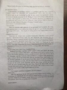
The classical treatment for excessive foreskin has long been surgical circumcision, with little breakthroughs in recent years. From October 2003 to February 2005, our hospital treated 52 cases of excessive foreskin using a minimally invasive surgical technique. Here we present the findings.

Materials and Methods

Clinical Data

The study included 52 patients, with ages ranging from 17 to 56 years and an average age of 38 years. Preoperative measurements of the penis in a flaccid state ranged from 2.5 cm to 10 cm. Of these, 40 were married and 12 were unmarried.

Surgical Technique
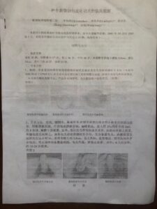
  1. Materials: The procedure utilizes a minimally invasive surgical ring invented by Mr. Shang Jianzhong, a special researcher at the Chinese Academy of Management Sciences (Patent No. 2003.ZL02 237969.X). The surgical ring is made from injection-molded polypropylene engineering plastic and consists of an inner and outer ring. The two rings are secured together using screws. The product comes in various sizes and is for one-time use in sterile packaging.

Attached Figure 1: Inner Ring of the Minimally Invasive Foreskin Cutter, Outer Ring of the Minimally Invasive Foreskin Cutter, Complete Minimally Invasive Foreskin Cutter (See insert for illustration).

原文插图

Methodology
  1. Preparation: Sterilization is performed, and a hole towel is laid out to expose the penis. A rubber band tourniquet is placed around the base of the penis to block venous return. A distended vein is then punctured, stagnant blood is aspirated, and 2ml of 2% lidocaine is injected. After waiting for 5 minutes, anesthesia is found to be highly satisfactory and complete.

  2. Ring Placement: An appropriately-sized surgical foreskin ring is chosen. The inner ring is first placed around the penis. The foreskin is then everted over the inner ring. If phimosis is present, a small incision is made on the dorsal side to fully expose the glans. The inner plate is retained up to 0.5 cm beyond the coronal sulcus, and the frenulum is left slightly longer, about 1.0 cm.

  3. Outer Ring and Cutting: The outer ring is then placed and screws are tightened. Excess foreskin protruding beyond the compression ring is trimmed. A sterile gauze strip is used to cover the wound, leaving the glans exposed. The tourniquet is then released, completing the surgery.

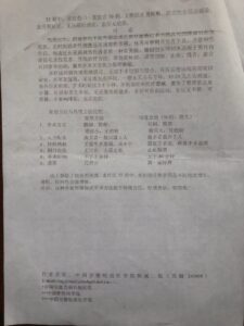
  4. Post-Operative Care: The ring is removed on the sixth day post-operation, and full recovery is generally achieved in approximately 15-20 days.

Attached Figure 2: Post-healing of Minimally Invasive Foreskin Surgery, Completion of Minimally Invasive Foreskin Surgery, Pre Minimally Invasive Foreskin Surgery (See above insert for illustration).

Results

Out of the 52 cases, primary wound healing was achieved in 50 cases post-operatively. In 2 cases, healing was delayed due to infection caused by engaging in sexual activity before the advised period. There were no long-term complications, and the healed wounds left no scars.

Discussion

Excessive foreskin length can lead to phimosis, where the coronal sulcus is not exposed, causing a buildup of secretions that cannot be eliminated, thereby leading to balanoposthitis. Long-term inflammation could even induce penile cancer. Phimosis can also result in poor penile development and impact sexual life. Excessive foreskin is a common issue plaguing male patients.

Minimally invasive foreskin ring resection is suitable for males with excessive foreskin and phimosis. Traditional treatment methods, such as full circumcision, involve cutting, hemostasis, and suturing, and often leave scars after healing; laser surgery also has drawbacks like thermal injury.

This innovative method breaks away from traditional approaches. It eliminates the need for surgical cutting and suturing. After the ring compresses the distal tissues, ischemia leads to tissue necrosis and eventual detachment, thus completing the circumcision. Generally, the ring is removed around the 6th day, and full recovery is achieved in approximately 15-20 days. The healed wounds leave no scars, and the surgery time is only 2-5 minutes. No additional medical equipment is needed, avoiding complicated hemostasis steps. The incidence of infection is low, no estrogen therapy is needed, and patients can move freely post-operatively. Daily life is not impacted; patients can bathe, urination is unaffected, and there are no complications.

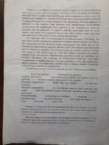
Comparison of New Method and Traditional Methods


New Method

Traditional (circumcision,laser) 

1、surgery

micro, convenient, no pain, no scars

invasive and complicated, with pains and scars

2、resources

one operator only

at least 2 operators needed

3、materials

no need for surgery tool

needs surgery tool in surgery room

4、bleeding

no bleeding

bleeding

5、procedure time

less than 5 min

more than 30 min

6、cost

low cost

more cost

In the 52 cases treated with this method, some patients experienced varying degrees of penile length and girth increase post-operatively, along with enhanced sexual function, due to the alleviation of the restrictions imposed by the foreskin.

Conclusion

This novel minimally invasive surgical approach is superior to traditional methods, with definite therapeutic effects. It is worthy of broader adoption.

原文插图


本文原载…….???


Originally published in "???" 90; 4(3):66 by Li Yangzhen, Li Mingjie, Shang Jianzhong, Wang Tong

【李名杰从医67年论文专辑】(电子版)

【李名杰从医67年论文专辑(英语电子版)】

Several improvement measures of circumcision

Surgical paper XIX

Several improvement measures of circumcision

Introduction

This paper outlines several innovative methods aimed at improving the effectiveness and safety of circumcision surgeries. The techniques include local venous anesthesia of the foreskin, bloodless surgery through arterial blockage, and the use of human hair as a suturing material.

1. Local Venous Anesthesia of the Foreskin

Procedure:

A rubber band is tied around the base of the penis to block venous return, causing the superficial veins of the penis to engorge. A fine needle is used to puncture the subcutaneous vein on the distal side of the penis, and 2 ml of 2% lidocaine is injected after aspirating stagnant blood.

Advantages:
  • Provides complete anesthesia.
  • Eliminates the risk of foreskin edema due to local anesthesia, thereby ensuring a more accurate resection line.

2. Bloodless Surgery via Blood Supply Blockage

Procedure:

The rubber band at the base of the penis is tightened, blocking the dorsal artery and creating a blood-free surgical field. This facilitates precise and complete cutting of the foreskin. Visible ends of severed blood vessels are first ligated, and then the tourniquet is released, ensuring complete hemostasis.

Advantages:
  • Creates a clear, blood-free surgical field.
  • Allows for precise cutting and complete hemostasis, aiding the surgical process.

3. Utilizing Human Hair for Suturing

Procedure:

Several strands of long female hair are cut and sterilized by soaking in a disinfectant solution for 5 minutes, followed by a saline rinse. The sterilized hair is then used as suture material.

Advantages:
  • Enables flexible suturing of both the inner and outer layers of the foreskin, ensuring a tight and accurate anastomosis.
  • Eliminates the need for the conventional 8-point suturing method, reducing contamination and facilitating healing.
  • Postoperative suture removal is not necessary as the hair naturally breaks and falls off within 7–10 days.

Results:

In a series of 30 cases using these techniques, there were no incidents of wound dehiscence or infection, and all cases achieved Grade I healing.

These improved circumcision techniques offer multiple benefits, including enhanced anesthesia, a cleaner surgical field, and easier postoperative care.

论文刊于《交通医学》90;4(3):66)???

Originally published in "Transportation Medicine Journal" 90; 4(3):66 by Li Mingjie, Changhang Hospital, 1979; 1:70

【李名杰从医67年论文专辑】(电子版)

【李名杰从医67年论文专辑(英语电子版)】

Subcutaneous heterotopic pancreas of abdominal wall

Surgical paper XIIX

Subcutaneous heterotopic pancreas of the abdominal wall

A Case Report

Introduction

Heterotopic pancreas is typically documented to occur only in internal organs, most commonly beneath the mucosa of the digestive tract. Clinically, it is rare to encounter. To date, there have been no reported cases of heterotopic pancreas located subcutaneously in the abdominal wall. We treated a case in our hospital, which was pathologically confirmed post-surgical resection. We present the report below.

Case Presentation

The patient is a 41-year-old male teacher admitted to our hospital on March 24, 1979, with the medical record number 794.

Approximately one year prior, he had a mass removed from his left lower abdomen at Hospital X. The pathological report identified the mass as a "desmoid tumor." Ten months post-surgery, a recurrent mass the size of a fingertip appeared at the same site, which was asymptomatic. In the past week, the mass suddenly enlarged to the size of an egg and became tender to touch.

On examination, the patient's general condition was normal, with no swollen superficial lymph nodes. A 5 cm oblique surgical scar was observed in the left lower abdomen. Below the scar, a 5x4x3 cm nodular, soft mass was palpable, with limited mobility and mild tenderness. There was no erythema or warmth. The white blood cell count was 5600/mm³, with 58% neutrophils and 42% lymphocytes.

Surgery was performed under local anesthesia immediately upon admission. The mass was located subcutaneously and above the muscle fascia. The boundary was ill-defined, and there was no capsule. The mass was lobulated and yellow in color. A sharp dissection was performed to excise the mass, followed by 24-hour subcutaneous drainage. The incision healed primarily, and the patient was discharged after a 9-day hospital stay. The pathological report confirmed "Heterotopic Pancreas in the Abdominal Wall's Fibrous Tissue" (Pathology No. 4686).

Postoperative Follow-Up

One week after discharge, the patient experienced subcutaneous swelling, pain, and fluctuation, although there was no redness or fever. Antibiotic treatment was administered by the local community hospital, leading to gradual resolution of the symptoms without the need for fluid aspiration. The patient returned to normal within a month.


Originally published in "Nanling Medical Journal" by Li Mingjie & He Jinxian, Department of Surgery, Nanling County Hospital, 1979; 1:70

【李名杰从医67年论文专辑】(电子版)

【李名杰从医67年论文专辑(英语电子版)】

Abdominal trauma

Surgical paper XIIIX

Abdominal trauma

Literature Review 

Introduction

Abdominal trauma, encompassing injuries to both the abdominal wall and internal viscera, is a prevalent medical issue. While the severity of the injury is undoubtedly a significant factor, early diagnosis and appropriate treatment are equally crucial. However, healthcare providers often encounter specific challenges and pitfalls, such as:

Common Pitfalls in Diagnosis and Treatment

  1. Unknown Medical History: The inability to obtain a valuable medical history due to patient coma or overlooked minor trauma.

  2. Subtle Early Symptoms: Lack of apparent signs in the immediate aftermath of the injury, making diagnosis challenging.

  3. Delayed Visceral Rupture: Instances of late-onset rupture that go unnoticed during the initial evaluation.

  4. Distracting Injuries: Concomitant severe injuries, such as cerebral or thoracic trauma, that divert attention away from abdominal injuries.

  5. Misdiagnosis Due to Spinal Involvement: Abdominal signs resulting from lower thoracic spine and chest injuries that may be mistaken for abdominal trauma.

  6. Conservative Approach: An overly cautious attitude, especially when the abdominal wall appears intact, can lead to missed diagnoses and lost opportunities for timely intervention.

Pathogenesis

abdominal trauma refers to injuries to the abdominal wall or internal organs, or both, caused by external force.

Mechanisms of Injury

  1. Direct Impact: Blunt force trauma to the abdomen.

  2. Indirect Impact: Falls from heights, seismic shocks, and other indirect forces.

  3. Non-penetrating Injury: Injuries resulting from vehicular accidents.

  4. Penetrating Injury: Wounds from knives, guns, or explosions.

Categories of Abdominal Viscera

  1. Solid Organs: Liver, spleen, kidney, pancreas.

  2. Cavitary Organs: Stomach, intestines, bladder, gall bladder.

  3. Structural Components: Mesentery, peritoneum, nerves, blood vessels.

Risks and Complications

The main risk of trauma to solid organs and stent structures is hemorrhage; the main risk for hollow organ trauma is the infection caused by the contents overflowing into the abdominal cavity, leading to peritonitis.

In fact, any visceral trauma can result in fatal hemorrhage. Liver and spleen injuries, in particular, can lead to long-term, persistent, and recurrent bleeding. This is because the blood vessel walls of these organs are thin, and the organs themselves are fragile and lack elasticity. Therefore, effective vessel constriction and blood clot formation may not occur. Moreover, once bleeding stops, due to the rich blood supply to these organs, a rise in blood pressure can wash away the hemostatic clot, leading to recurrent bleeding.

The likelihood of hollow organ injury is related to the amount of contents it holds (those with a full stomach are more susceptible to injury); it is related to the degree of fixation (the distal and proximal ends of the small intestine, and the hepatic and splenic flexures of the colon are vulnerable); and it is related to pre-existing conditions (those with prior illnesses are more susceptible to injury).

The nature of substances that irritate the peritoneum determines its pathological changes and abdominal signs:

  1. Gastrointestinal bacteria increase from top to bottom, such as in the case of fecal peritonitis due to colon injury, which has a very strong infectious nature.

  2. The chemical irritability of the gastrointestinal tract decreases from top to bottom, such as in the case of chemical peritonitis caused by perforations in the stomach or duodenum, which can be very severe.

  3. Bile and urine may or may not contain bacteria, but they are chemical mixtures that can lead to peritonitis, albeit a bit later in time.

  4. Blood without bacteria causes minimal irritation to the peritoneum, so the reaction to hemorrhagic peritonitis is usually mild.

  5. Gas irritation of the peritoneum can also produce symptoms.

Regardless of the nature of the material that spills into the abdominal cavity, it often follows its natural drainage route with the peritoneal exudate it produces, leading to the paracolic gutter and the pelvic floor, resulting in intestinal paralysis and abdominal distension.

Symptoms and Signs

  1. Fainting or Shock:

    • Abdominal wall contusions are rare, but visceral injuries are common. Substantial visceral organ rupture can cause massive bleeding and can be extremely dangerous. Damage to major blood vessels can be immediately fatal and beyond rescue. Peritonitis due to the perforation of hollow organs leads to toxic shock.
  2. Abdominal Pain:

    • Contusions to the abdominal wall are localized and minor; visceral injuries are extensive and persistent.
  3. Abdominal Distension:

    • Progressive worsening is a sign of internal bleeding and peritonitis, with inhibited abdominal breathing. However, attention must also be paid to intestinal motility inhibition caused by retroperitoneal injuries and hematomas that stimulate the abdominal plexus.
  4. Abdominal Tenderness:

    • Point muscle guarding, tenderness, and rebound tenderness can often indicate the site of injury in the early stages. In the later stages, it becomes generalized and is often corroborative evidence of visceral injury.
  5. Nausea and Vomiting:

    • Reflexive in the early stages and due to backflow in the later stages.
  6. Liver Dullness Boundary:

    • Abdominal gas distension can cause it to shrink, and a free air layer can make it disappear. After liver rupture, the dullness boundary expands.
  7. Shifting Dullness:

    • Often a significant basis for abdominal hemorrhage or effusion.
  8. Intestinal Motility:

    • Reflexively weakened in the early stages, leading to an "inactive abdomen" caused by inflammation in the later stages.

Diagnosis

The central issues in diagnosing abdominal trauma are to clarify:

  1. Is it a simple abdominal wall injury, or is there also visceral injury?
  2. Which organ(s) are injured, and is it a single or multiple injuries?
  3. What is the volume and rate of internal bleeding, and has it stopped or is it ongoing?

A detailed medical history should be obtained, including the magnitude and direction of the force, posture at the time of injury, and subsequent reactions such as sudden sharp pain, fainting, and vomiting. Physical examination should include evaluation of muscle guarding, the scope of tenderness, dullness and tympanic areas, and their changes. Body temperature, pulse, respiration, blood pressure, blood and urine tests, X-rays, and ultrasound should be measured. The use of analgesics like morphine should be avoided during observation, except when a diagnosis has been confirmed and surgery is planned. If necessary, exploratory laparotomy should be performed; it's better to act than to miss the opportunity, even if no significant findings are obtained.

  1. Blood Count: An elevated white blood cell count in the early stage is a physiological response to trauma. Some believe that internal bleeding can produce a large amount of hematopoietic stimulants, leading to an elevated white blood cell count when absorbed by the peritoneum. Peritonitis naturally causes elevated white blood cells and a left shift in neutrophils. Internal bleeding presents as anemia.

  2. Hematuria: A strong basis for urological injuries and an effective indicator for monitoring the outcome of the injury.

  3. X-Ray: Free air under the diaphragm is generally considered to have only a 50% positive rate, and attention should be paid to it. Comparison of the diaphragmatic height and movement on both sides can aid in the diagnosis of liver and spleen injuries.

  4. Abdominal Paracentesis: Highly valuable. The characteristics and state of the extracted fluid can often confirm the diagnosis. Because a positive result requires more than 200 ml of fluid in the abdominal cavity, some use lavage to increase the positivity rate. If unsuccessful, change the needle direction and depth or reattempt after an interval. Besides macroscopic observation, amylase quantification and smear tests can be done on the puncture fluid. The presence of multiple types of bacteria without pus cells or the presence of parasitic eggs supports the hypothesis of accidental intestinal entry.

  5. Rectal Examination and Posterior Fornix Puncture in Married Women: These can be included as routine examinations to help with early diagnosis.

Management and Treatment

  1. Shock Management: For patients in shock, use a slight incline position and elevate both legs (to facilitate breathing and venous return). Measures like warming, fluid resuscitation, blood transfusion, oxygen administration, and antibiotics are essential. Given the peritoneum's strong anti-infective ability, fatalities due to infection are far less common than those due to shock or bleeding. Therefore, establishing an open vein and rapidly restoring effective blood volume is of utmost importance.

  2. Surgical Intervention: Surgery for abdominal injuries should ideally be performed within 6 hours. If anti-shock measures are ineffective, forced surgery should be performed to eliminate the source of the problem as one of the life-saving measures.

  3. Surgical Incision: The location of the incision is determined based on the estimated site of injury. The principle is to make the incision as close and convenient as possible to the affected area. A midline vertical incision is generally used, as it allows for quick entry and is easily extendable. Before closing the abdomen, it's crucial to thoroughly wash and aspirate the abdominal cavity. For contamination with bile, pancreas, feces, or urine, drainage should be implemented.

Specific Injuries and Treatments

  1. Spleen Rupture: Standard procedure is removal. Some recent studies advocate for repair in minor injuries to preserve spleen function. Autotransfusion of the patient's own blood can be done without anticoagulants, saving resources and avoiding citrate toxicity.

  2. Small Intestine Rupture: Repairs or resection and anastomosis are the go-to approaches. Externalization of the intestine is less preferred.

  3. Liver Injury:

    • Type of Injury: Most liver injuries involve a laceration of the liver tissue, which largely remains intact due to the resiliency of its blood vessels.
    • Surgical Interventions: During surgery, the focus is on debridement and individual ligation of severed blood and bile ducts. Further harm to the liver should be avoided. Full-layer mattress sutures are generally used, and the omentum is often laid over the sutured area. Sometimes, omentum or muscle can be used for packing. Gauze packing should be avoided.
    • Limited Crush Injuries: For localized crush injuries, partial hepatectomy is recommended.
    • Control of Bleeding: If there is profuse bleeding during surgery, temporary occlusion of the hepatic portal can control it (up to 15 minutes at room temperature is allowed). If bleeding can't be controlled, ligation of the hepatic artery often works immediately.
    • Post-Operative Considerations: Adequate drainage below the liver is crucial, often with the additional step of controlled decompression of the common bile duct.

    Suturing and Complications:

    • Suture Material: Both intestinal thread and silk thread are acceptable, although the author believes that No. 4 silk thread is preferable.
    • Tightness of Ligations: Ligations should not be too tight; they should be just tight enough to control bleeding. Over-tightening can lead to complications like hepatic necrosis, delayed bleeding, or bile leakage.
    • Anecdotal Experience: The author recounts a case of postoperative massive biliary bleeding on Day 21, which required another surgery to ligate the intrinsic hepatic artery.

    Blood Transfusion:

    • Caution with Bile-Contaminated Blood: Normally, blood contaminated with bile should not be transfused back. However, an anecdotal case saw 1500 mL transfused without adverse effects in a rural setting. It's generally considered safe to transfuse one's own blood back if there are no injuries to the extrahepatic biliary tract.
  4. Colon Injury: Early repair is recommended; late-stage injuries may require externalization and later resection and anastomosis.

  5. Mesenteric Tear: Repair and hemostasis are essential. If blood supply to the intestines is compromised, resection may be needed.

  6. Pancreatic Injury: Small tears can be sutured, and the pancreatic duct can be ligated if damaged. For injuries to the head of the pancreas, reimplantation is necessary to establish a new pancreatic-intestinal pathway.

  7. Stomach and Duodenal Injuries: Repair is standard. Special attention should be given to retroperitoneal injuries of the duodenum.

  8. Extrahepatic Biliary Tract Injury: Repair, fistula creation, and external drainage are options.

  9. Kidney Injury: Generally conservative treatment under close observation is sufficient due to the kidney's high regenerative capacity.

    10. Extrarenal Urinary Tract Injury: Injuries to the ureter are rare. Bladder injuries are more common and often associated with pelvic fractures. Repair, fistula creation, and drainage are usual treatments.

    11. Diaphragm Injury: More commonly seen on the left side, resulting in diaphragmatic hernia and bleeding. High mortality rates necessitate early thoracic repair.


    12. Retroperitoneal Major Vascular Injury
    : Repair is generally performed unless immediate intervention is not possible. For injuries to the inferior vena cava below the renal veins, ligation is an option.

    • General Treatment: Unless the injury is too severe to be managed emergently, the usual approach is to repair the damaged vessel.
    • Below the Level of the Renal Vein: Injuries to the inferior vena cava below the level of the renal veins can generally be managed by ligation and cutting off the damaged segment.
    • Above the Level of the Renal Vein: For injuries to the inferior vena cava above the level of the renal veins, if repair is not possible, some advocate for ligation. However, this necessitates the removal of the right kidney. The left kidney can rely on collateral circulation (e.g., adrenal vein) and should be unaffected. However, this approach could lead to insufficient venous return to the heart.

    Discussion

    Early Diagnosis:

    Determining whether the injury is confined to the abdominal wall or involves internal organs is crucial for making timely decisions about surgical intervention and life-saving measures. A simple contusion of the abdominal wall can also lead to symptoms like abdominal pain, muscular guarding, and localized tenderness due to muscle bruising and bleeding, which can irritate the peritoneum. These symptoms may even be accompanied by nausea and vomiting. On the other hand, early signs of internal organ damage can be subtle due to minimal initial bleeding and leakage, which may be diluted by intraperitoneal fluid. This makes it easy to confuse the two types of injuries.

    Abdominal Wall Contusion:

    In cases of abdominal wall contusion, the abdominal pain is usually mild, muscular guarding is localized, and there may be subcutaneous bruising and soft tissue swelling. Generally, systemic disturbance is minimal, and shock is unlikely.

    Solid Organ Rupture:

    For solid organs, except for the liver and gallbladder, the symptoms of peritoneal irritation are generally less severe compared to hollow organ injuries. While pain may be felt throughout the abdomen, it is most prominent in the area where the damaged organ is located.  The following points can assist in the diagnosis:

    1. Indicators of Internal Bleeding: After trauma to the lower chest and upper abdominal area, there may be signs of internal bleeding, such as an increased pulse rate, a drop in blood pressure, progressive anemia, and the absence of abdominal breathing.

    2. Pain Radiating from the Ribcage: Abdominal pain may be most pronounced in the costal margins and may radiate to the shoulders, back, and waist.

    3. Persistent Upper Abdominal Pain: After the injury, pain in the upper abdomen persists and may be accompanied by localized tenderness, muscular guarding, abdominal distension, and reduced bowel sounds.

    4. Changes in Liver or Spleen Percussion: On X-ray, there may be an enlargement of the liver or spleen shadow, elevation and reduced movement of the diaphragm on the injured side, and disappearance of the psoas muscle shadow.

    5. Presence of Fixed or Shifting Dullness: During abdominal paracentesis, non-clotting dark red blood may be observed. This is due to defibrinated blood, which is also associated with the action of fibrinolysin.

Diagnosis of Hollow Organ Perforation:

Perforation of hollow organs often leads to peritonitis at an early stage, characterized by severe abdominal pain that makes early detection easier. The following points can assist in diagnosis:

  1. Severe Abdominal Pain Lasting Over 4 Hours: This is accompanied by:

    • Nausea and bilious vomiting
    • Increased pulse rate
    • Fixed tenderness upon palpation
    • An expanding area of muscular guarding
    • Elevated white blood cell count with a leftward shift in differential
    • Weakened or absent abdominal breathing
  2. Anemia Not Prominent but Rapid Pulse and Restlessness: Even if anemia is not obvious, a fast pulse rate and symptoms of restlessness or agitation may be present.

  3. Disappearance of Liver Dullness: An X-ray may show the presence of free air under the diaphragm, indicating perforation.

  4. Abdominal Paracentesis Shows Turbid Fluid: Microscopic examination confirms the presence of pathogens, indicating infection.

Early Diagnosis and Treatment:

For patients with multiple injuries, if there is even one reliable indicator, early exploratory laparotomy is advised.

Conditions for Conservative Treatment:

  1. Injury is confined to the abdominal wall.
  2. Although there's internal bleeding, it has stopped and there are no signs of hollow organ perforation; vital signs remain stable.
  3. General condition is still good one or two days post-injury, and any internal infection is localized.
  4. Mild kidney injuries where the hematuria gradually decreases and pain subsides.

Anesthesia:

Complete anesthesia is required to relax the abdominal muscles, facilitating exploration and cleaning of the abdominal cavity. Our majority of cases have safely and satisfactorily used continuous epidural anesthesia, but it should be administered in small, slow doses. Note that the required dosage for these patients is generally less than for typical patients.

Exploration:

Pay attention to the existence of compound injuries to avoid overlooking any. Check retroperitoneal organs like the duodenum, colon, and the bare area of the liver. The omentum often points to the location of the pathology.

Suggested Order of Exploration:

Spleen, liver, pancreas, diaphragm, base of the stomach, bile ducts, duodenum, mesentery, omentum, small intestine (from the jejunum to the ileocecal region), large intestine (note the hepatic and splenic flexures), retroperitoneal organs like kidneys and major blood vessels, and pelvic organs.

Drainage:

The decision to drain is based on the timing of the injury, the degree of infection, and the quantity and quality of the intra-abdominal fluid. For early abdominal contamination, thorough washing is the main approach, supplemented by drainage. Drainage is necessary for liver, gallbladder, pancreas, and intestinal injuries.

Prognosis:

Early death is usually due to severe compound injuries, traumatic shock, and major bleeding. Late death often occurs from generalized peritonitis, toxic shock, and water and electrolyte imbalances. The cure rate for surgical cases can reach around 90%.

References:

  1. Huang Jiaqi, "Surgery," People's Health Publishing House, P: 497, 1964.
  2. Gao Xianming, "Abdominal Trauma," Journal of Surgery, 6: 468, 1951.
  3. Jiang Kegou, "Diagnosis and Treatment of Abdominal Trauma," Journal of Surgery, 6: 376, 1951.
  4. Huang Wen, "Clinical Analysis of 98 Cases of Abdominal Contusion," Chinese Journal of Surgery, 4: 370, 1960.
  5. Gong Songnan, "Closed Abdominal Injury (with analysis of 220 cases)," Chinese Journal of Surgery, 15: (2) 96, 1977.


Originally published in "Nanling Medical Journal" by Li Mingjie, Department of Surgery, Nanling County Hospital, 1979; 59-63.


【李名杰从医67年论文专辑】(电子版)

【李名杰从医67年论文专辑(英语电子版)】

A case of plastic tube foreign body in bladder

Surgical paper XVI

A case of plastic tube foreign body in the bladder

Patient Information

  • Gender: Male
  • Age: 20
  • Occupation: Farmer
  • Hospital Admission Number: 16318

History

On April 5, 1990, out of curiosity and playfulness, the patient self-inserted a 35-cm hollow plastic tube designed for hair-tying into his bladder via the urethra and was unable to remove it. He subsequently developed symptoms of lower urinary tract irritation, including frequent urination, urgency, and painful urination. Local hospital urine tests revealed pyuria (++), but a plain film of the bladder was reported as negative. Due to the concealment of this medical history, treatments for cystitis were ineffective. The patient was admitted to the hospital on December 3, 1990.

Physical Examination

General conditions were normal. Urinalysis showed red blood cells (++) and pyuria (x10). B-mode ultrasound revealed a hyperechoic mass within the bladder. A plain film of the bladder showed a circled mass (Figure 1). The diagnosis was a foreign body in the bladder accompanied by stone formation.

Surgical Intervention

On an unspecified day in December 1990, the patient underwent a cystotomy under continuous epidural anesthesia. A solid foreign body, measuring 2×2.5×3.0 cm and weighing 5.5 grams, was removed (Figure 2). It consisted of five loops of the plastic tube, folded upon themselves, with extensive urine salt deposition.

Insights and Reflections

This case underscores the importance of a detailed medical history for accurate diagnosis. After the operation, we conducted a simulation using a hollow plastic tube similar to the one in the patient's case and found that if the conditions are right and the films are read carefully, the foreign body could indeed be identified. However, in the early stages of this case, the local hospital misinterpreted a bladder plain film as negative and incorrectly treated the patient for cystitis. This not only prolonged the symptoms but also led to the deposition of urine salts around the foreign body, turning it into a calcified mass. Essentially, this became a case of secondary bladder calculus.

Interestingly, because the foreign body served as the core around which salts aggregated, it did not readily cause obstruction during urination. Thus, symptoms like interrupted urine flow or "staccato" urination were absent. Instead, the patient continued to experience pain and symptoms of bladder irritation. The correct diagnosis was eventually made based on the patient’s medical history, B-mode ultrasound, and X-ray examinations.

Given the specific circumstances of this case, attempting to break and remove the stone via the urethra seemed implausible and would likely result in remnants. Complete surgical removal of the mass proved to be the most effective treatment approach.


(1991/10/05), Changhang Hospital, Li Mingjie & Shi Lianghui

【李名杰从医67年论文专辑】(电子版)

【李名杰从医67年论文专辑(英语电子版)】

Recurrent stones in common bile duct with suture as core

Surgical paper XV

Recurrent stones in common bile duct with suture as core 

A report of 6 cases

Introduction

Between 1975 and 1980, we reviewed a group of 70 patients who had undergone reoperations for recurrent cholelithiasis, representing 19% (70/368) of cholelithiasis surgeries during the same period. Of these 70 cases, six had recurrent stones formed around the surgical suture used in their previous operations. This report aims to discuss these specific six cases due to their implications for improving surgical techniques.

Case Summaries

Case 1

  • Gender: Male
  • Age: 37
  • Hospital No.: 1047
  • Date of Admission: February 13, 1976
  • Medical History: Underwent cholecystectomy, choledocholithotomy, and T-tube external drainage at our hospital a year prior.
  • Symptoms: Began experiencing frequent right upper abdominal pain and occasional fever six months after discharge.
  • Intraoperative Findings: Discovered a 2cm diameter muddy stone mass formed around a surgical suture in the common bile duct.
  • Outcome: Stone removed, T-tube placed, and the patient was discharged 14 days post-op. No recurrence at three-year follow-up.

Case 2

  • Gender: Female
  • Age: 28
  • Hospital No.: 4893
  • Date of Admission: October 6, 1977
  • Medical History: Had cholecystectomy, left hepatic lobectomy, and T-tube external drainage two years earlier.
  • Symptoms: Upper right abdominal pain, fever, and jaundice half a month prior.
  • Intraoperative Findings: Two pieces of 7cm and 4cm sutures were found in the common bile duct, around which loose stone masses of 3cm and 2cm in diameter had formed.
  • Outcome: Stones removed, patient was hospitalized for 15 days and discharged. However, the patient died a year later due to intrahepatic stones and severe infection, leading to shock.

Case 3

  • Gender: Female
  • Age: 52
  • Hospital No.: 5105
  • Date of Admission: October 20, 1977
  • Medical History: Cholecystectomy and common bile duct T-tube drainage 14 months ago.
  • Symptoms: Recurrence of symptoms 8 months post-op, leading to admission 10 hours after the onset.
  • Intraoperative Findings: A 4x3x3 cm sand-mud stone mass was found, with the suture from the previous surgery serving as the core.
  • Outcome: The patient died of toxic shock 10 hours post-operation.

Note: Three additional cases had similar intraoperative findings and outcomes. All were successfully treated and had no recurrence after a follow-up period of 1 to 3 years.

Discussion

The recurrence rate of primary hepatobiliary pigmented stones after surgery is high, reaching 19% according to our data. The fundamental cause is metabolic dysfunction in bile composition. However, improper surgical procedures or the retention of foreign objects can also lead to early recurrence of stones. In this report, all six cases of recurrent stones had sutures from previous surgeries as their core. The absence of these sutures could have potentially prevented the recurrence or at least recent postoperative recurrence. Case 3 in our series tragically passed away due to this issue, serving as a serious lesson.

Although we have only identified six such cases, the actual number may be higher. Not every stone removed in reoperations is crushed for examination. Therefore, it is possible that the actual number of such cases is higher. Furthermore, in most reoperations, the original sutures from previous surgeries on the common bile duct were not found. We speculate that once these sutures penetrate into the lumen, the body's natural rejection mechanisms could expel them into the intestine through the common bile duct, thereby averting harm. However, before being expelled into the intestine, could these sutures serve as a nucleus for stone formation or even cause symptoms? This warrants further investigation. It might also be one of the reasons contributing to the so-called "post-cholecystectomy syndrome," another aspect deserving attention. Further systematic endoscopic or radiographic studies are needed to draw scientific conclusions.

In our hospital, the common practice for suturing the common bile duct incision involves using non-absorbable No.0 or No.1 silk threads for full-layer continuous suturing, starting from the upper edge of the incision and moving downwards. While this method ensures tight closure and is time-efficient, it has a downside. During the removal of the T-tube, part of the duct wall is inevitably damaged, exposing the suture to the lumen. This can lead to a natural rejection response, causing the suture to adhere to bile sediments before being fully expelled into the intestine, sometimes leading to symptoms. Therefore, we recommend abandoning this suturing method and instead using thinner sutures for submucosal interrupted sutures to prevent the suture from being exposed to the lumen.

Case 3 demonstrated that even the thicker sutures used for the hepatic section can fall into the lumen, illuminating the body's natural rejection instinct. This is a rare but instructive instance.

References:

Li Mingjie: Domestic Medical Abstracts Surgery Volume. Nanning, Guangxi Medical Institute 1981; 39 (0161)


Originally published in the Proceedings of the Second Annual Surgical Conference of Anhui Province, 1988; 87; presented at he Yangtze River Full-Line Biliary Surgery Seminar, No. 23. (1987/02/24), Changhang Hospital, Li Mingjie

【李名杰从医67年论文专辑】(电子版)

【李名杰从医67年论文专辑(英语电子版)】

Primary repair of congenital omphalocele 

Surgical paper XIV

Successful primary repair of congenital omphalocele 

A Case Report

Case Presentation

The patient, a male newborn named Shao, was born at home on May 14, 1983. He was brought to the emergency department four hours post-birth due to the presence of an omphalocele, a condition where part of his abdominal organs were protruding out of the abdomen, enclosed in a transparent membranous sac.

Clinical Examination

Upon examination, the infant weighed 3010 grams and had no other associated congenital malformations. The umbilical cord was ligated at a distance of 10 cm. The transparent sac contained portions of the liver, the majority of the stomach, and sections of the small and transverse intestines. There were no signs of strangulation or vascular compromise. The sac was intact and not discolored, and the abdominal wall defect at the umbilicus measured 6 cm (Figures 1 and 2).

Surgical Intervention

The surgery was performed under local anesthesia. The omphalocele sac was excised, and the skin edges were trimmed and dissected laterally. Successful herniation of the protruding organs was achieved, followed by layered suturing (Figure 3). The patient experienced neither respiratory distress nor circulatory complications. He passed gas and stool on the evening following surgery, and sutures were removed as scheduled. The wound healed without complications.

Follow-up

One-and-a-half years post-surgery, the child displayed normal development, stable gait, and no intellectual disabilities. He had begun to recognize and call out to people.

Discussion

Congenital omphalocele is an embryonic developmental disorder resulting from the failure of synchronous development between the abdominal cavity and viscera. Approximately 40% of cases present with additional congenital malformations. The condition is relatively rare, occurring in 1 in 7,000 births. The literature contains limited case reports, with one study reporting only 22 instances. Delayed surgical intervention can result in desiccation, necrosis, and rupture of the omphalocele sac, exposing the organs to infection and significantly increasing mortality risk. The surgical approach, either primary or staged repair, depends on the size of the omphalocele and the developmental status of the abdominal cavity. Our case, although categorized as a "giant" omphalocele due to the 6 cm abdominal wall defect and involvement of multiple organs, was successfully managed through primary repair owing to the absence of other deformities and favorable overall development.


References

  • She Yaxiong: Pediatric Surgery. 1979; p. 296

Figures: Embryonal Omphalocele

  • Figure 1: External appearance of the omphalocele with a 6 cm abdominal wall defect at the umbilical pedicle.

  • Figure 2: Organs involved in the omphalocele: liver, stomach, small intestine, and transverse colon.

  • Figure 3: Appearance following successful primary repair.


1988/04/01
Changhang Hospital, Li Mingjie

【李名杰从医67年论文专辑】(电子版)

【李名杰从医67年论文专辑(英语电子版)】

Adult retroperitoneal teratoma infection complicated with chronic purulent fistula

Surgical paper XII

Adult retroperitoneal teratoma infection complicated with chronic purulent fistula 

A Case Report

Introduction

A 53-year-old female patient presented with a history of left waist swelling, pain, fever, and pus discharge that occurred 12 years ago and reportedly "self-healed" within a few months. Six years prior to the current consultation, she developed an abscess on the medial aspect of her left thigh, which was incised to create a fistula but never fully healed.

Clinical Examination

Upon examination, the patient appeared to be suffering from chronic consumptive anemia. She had purulent fistulas in both the right lumbar and left femoral regions, discharging yellowish fluid and some pus. Radiographic evaluations showed no abnormalities in the spine, pelvis, or hip joints. However, the shadow of the left psoas muscle appeared indistinct. No evidence of an intestinal fistula was found through contrast studies. A 4.5 cm irregular residual barium shadow was noted near the second and third lumbar vertebrae, appearing potato-like. Biopsy of the fistula tissue revealed inflammatory granulation.

Surgical Findings

Immediate surgical exploration was carried out, confirming that the fistula tract led into the left psoas muscle. Upon injecting methylene blue into the fistula and subsequently incising the muscle, thin pus was drained. The abscess cavity was spindle-shaped, measuring approximately 20x5x4 cm. Tracking the cavity upwards led to the discovery of a 6x5x4 cm encapsulated mass near the second and third lumbar vertebrae. The mass was easily excised. It was adherent to and penetrated the psoas major muscle. The intramuscular abscess was fully incised, its inner wall scraped clean of granulation tissue, washed, and hemostasis was achieved through compression.

Pathological Diagnosis

The pathological examination confirmed a benign cystic teratoma complicated by infection, leading to the formation of a left psoas muscle abscess and an external fistula.


This article was originally published in Journal of Transportation Medicine,1993;Vol.7, fourth (368)
Changhang Hospital, Li Mingjie

【李名杰从医67年论文专辑】(电子版)

【李名杰从医67年论文专辑(英语电子版)】

Lighter foreign body in stomach

Surgical paper XIII

Ingested lighter as a foreign body in the stomach

A Case Report

Case presentation

A 22-year-old male, generally in good health, intentionally ingested a wire resistance lighter two months prior to admission. He manually forced the object through his pharynx, facilitating its smooth passage into the stomach without experiencing obstruction, bleeding, or esophageal injury. Despite consuming a large quantity of leeks that evening, the foreign body was not naturally expelled and he experienced no immediate discomfort. One month later, he began experiencing abdominal pain on an empty stomach, which was alleviated by eating. An abdominal X-ray at our institution confirmed the object's retention in the stomach. Surgical intervention was deferred until two months later, given the absence of acute symptoms.

Clinical Examination

Upon physical examination, the patient appeared generally well, with no upper abdominal tenderness. Abdominal X-ray and B-type ultrasonic gastric perfusion examination confirmed the presence of the foreign body within the stomach.

Surgical Procedure

On July 4, 1991, under epidural anesthesia, a 3.5 cm gastric incision was made. The gastric mucosa appeared slightly congested, but there were no signs of ulceration or hyperplasia. The foreign object, not adhered to the gastric wall, was successfully retrieved under direct visualization and the stomach was subsequently repaired. The postoperative course was uneventful, with sutures removed on day seven.

Pathological Findings

The retrieved foreign body was identified as a wire resistance lighter, measuring 7x3x1.5 cm and weighing 18 grams. It was composed of a blend of plastic and metal. Erosion was evident on its metal shell, and part of the plastic had been stripped away.

Discussion

The smooth passage of a rigid foreign body with a 3 cm transverse diameter through the pharynx and esophagus without injury is notable. However, the object remained in the stomach for an extended period, unable to pass through the pylorus. Stomach acid had corroded both the metal and plastic components of the lighter, although it remained largely intact due to its composite nature. Endoscopic retrieval was considered but deemed risky due to the potential for iatrogenic injury. Surgical removal proved to be a safe and effective approach, with a favorable prognosis confirmed at a three-month follow-up.


1991/10/15
Changhang Hospital, Li Mingjie & Wang Yisheng

【李名杰从医67年论文专辑】(电子版)

【李名杰从医67年论文专辑(英语电子版)】

Misdiagnosis of subacute perforated peritonitis in gastric malignant lymphoma

Surgical paper XI

Misdiagnosis of subacute perforated peritonitis in gastric malignant lymphoma 

A Case Report

Case Presentation

A 71-year-old female patient, with the medical record number 13520, experienced intermittent periumbilical abdominal pain for 10 days, without accompanying diarrhea, hematochezia, or fever. She was admitted to the Affiliated Hospital of Anhui Medical University on March 22, 1987, with severe colic in the right lower abdomen and vomiting lasting for two days. She was initially suspected to have appendiceal perforation.

Clinical Examination

Upon admission, the patient had a temperature of 35.5°C, a pulse rate of 84 beats/min, and a blood pressure of 120/80 mmHg. She appeared acutely dehydrated with general nutrition. No lymphadenopathy or jaundice was observed. Physical examination revealed tenderness and rebound tenderness throughout the abdomen, especially in the right lower quadrant. Laboratory tests showed decreased hemoglobin levels (9 g/mm3), RBC count of 3.8 million/mm3, and a WBC count of 19,700/mm3 with 91% neutrophils and 9% lymphocytes. Urine amylase was 16 units/Winsler.

Surgical Findings

The patient underwent emergency surgery on the day of admission for suspected acute appendicitis with peritonitis. During the procedure, a small amount of intra-abdominal exudate was found, but no obvious purulent fluid was observed. The appendix, gallbladder, liver, spleen, and pancreas appeared normal. However, a 5x6 cm mass was discovered on the anterior wall of the gastric antrum, with a soft texture and focal necrosis at its center.

Histopathology and Postoperative Care

Histopathological examination confirmed the diagnosis of malignant lymphoma of the gastric antrum. The patient was discharged after 12 days of hospitalization and showed no signs of recurrence during the one-year follow-up.

Discussion

Gastric malignant lymphoma accounts for 80% of gastric sarcomas and often presents with insidious and non-specific symptoms, making preoperative diagnosis challenging—only 10% are accurately diagnosed before surgery. These tumors may result in peritonitis due to necrosis and exudation, often misleadingly presenting as appendicitis.

For patients with a prolonged history of symptoms but no localized inflammatory mass, the possibility of this rare condition should be considered. Excision of sufficient gastric tissue and omentum generally leads to a more favorable prognosis than gastric cancer, with a 5-year survival rate up to 50%. In this case, the patient remained symptom-free at the one-year follow-up.


References

Waltar LJ, et al. Cancer Management. New York, 1977; p.269.


This article was originally published in Proceedings of First Health Conference of the Yangtze River Shipping Company,1988;4:1
Changhang Hospital, Li Mingjie

【李名杰从医67年论文专辑】(电子版)

【李名杰从医67年论文专辑(英语电子版)】

Diagnosis and treatment of close duodenal retroperitoneal injury 

Surgical paper X

Diagnosis and treatment of close duodenal retroperitoneal injury

ABSTRACT

Closed duodenal injuries represent a unique and severe subtype of intra-abdominal trauma characterized by low incidence but high mortality rates. This study discusses six instances of retroperitoneal duodenal injuries, reporting a successful treatment outcome in four cases and fatalities in the remaining two. Such injuries are particularly elusive to early diagnosis due to their retroperitoneal location, which often results in the absence of overt symptoms and signs associated with hollow organ perforation. Therefore, clinicians must exercise heightened vigilance, carry out meticulous and ongoing dynamic monitoring, and seek robust diagnostic evidence to expedite surgical intervention. Given the specialized anatomical and physiological characteristics of the duodenum, the treatment approaches diverge significantly from those employed for other visceral injuries. This makes surgical choices pivotal to the prognosis. The study finds that comprehensive duodenal decompression and diverticularization techniques are dependable. The Berne procedure is particularly recommended for its efficacy in drainage and infection control, supplemented by requisite supportive care.

Keywords:
Retroperitoneal Duodenal Injury, Diverticularization, Berne Procedure

The duodenum, located deep within the posterior abdominal wall, is less frequently subjected to injury, making up only 3–5% of closed abdominal injuries and 10% of gastrointestinal injuries [1]. When a rupture occurs within the peritoneal cavity, it typically manifests quickly with signs of peritonitis, much like other hollow organ perforations. This draws immediate clinical attention, leading to timely surgical intervention. However, when the injury is confined to the retroperitoneal region, the leakage of intestinal fluids is concealed within the retroperitoneal space. This presents a diagnostic challenge as it lacks overt symptoms or positive physical signs, leading to delayed diagnosis and treatment. Consequently, the mortality rate for such injuries skyrockets to between 30–60% [2], posing a significant clinical conundrum.

Our hospital has treated 258 cases of closed abdominal injuries over the years, among which 8 involved duodenal injuries. Of these, 6 cases (2.3%) were retroperitoneal injuries. Each presented unique difficulties, resulting in delayed surgical interventions. Here, we dissect the complexities, experiences, and lessons gleaned from these cases.

Clinical Data

All patients in this study were male, ranging in age from 17 to 45 years. Of these, four sustained injuries to the descending part of the duodenum, while the remaining two had injuries in the transverse section. Two patients sought medical attention within four hours post-injury, and the other four within 24 hours. Associated injuries included one case each of liver trauma, inferior vena cava damage, mesenteric vascular injury, and splenic rupture. Additionally, two cases presented with isolated duodenal retroperitoneal injuries. The causes of injuries were varied: two resulted from falls, one from a blunt force injury by a wooden stick, and three from motor vehicle accidents.

As for the timing of surgical intervention, two patients underwent surgery within 24 hours post-injury due to concomitant severe intra-abdominal bleeding. The remaining patients were operated on between 24 and 48 hours post-injury, as positive abdominal signs progressively manifested.

Two patients received a straightforward repair followed by intraperitoneal drainage; however, both cases had unfortunate outcomes. One succumbed to hemorrhagic shock six hours post-surgery, and the other passed away on the 4th and 8th postoperative days due to complications from an intestinal fistula and subsequent infection and electrolyte imbalances, respectively.

The remaining four patients underwent a more complex surgical approach incorporating the Berne-like technique [3]. This involved duodenal and common bile duct fistulization, along with gastric-jejunal anastomosis following gastric antral resection. All four of these patients successfully recovered post-surgery.

Discussion

2.1 Mechanism of Injury

The injury mechanism is often a consequence of blunt trauma or inertial decompression, leading to a sudden shift in intra-abdominal pressure. This forces the duodenum against the spine and induces pyloric spasms, dramatically increasing intestinal pressure. Both internal and external bidirectional shearing forces act upon the frail and fixed duodenal wall, causing it to rupture.

2.2 Pathological Underpinnings of Duodenal Retroperitoneal Injury

In the early stages post-injury, leaked fluids accumulate locally in the ruptured area, manifesting few systemic symptoms and remaining largely undetectable. In our cohort, two cases featured isolated retroperitoneal injuries in the descending duodenum; surprisingly, these patients were ambulatory post-admission, experiencing only lower back discomfort. Symptoms generally worsened after 24 hours. A startling 80% of such cases are not definitively diagnosed preoperatively [4]. The leaking digestive fluids contain a myriad of components like hydrochloric acid, bile salts, cholesterol, and digestive enzymes, among others. These substances cause chemical irritation, autodigestion, and infection, leading to a cascade of complications, including severe inflammation, edema, necrosis, and multiple organ failure.

2.3 Diagnostic Key Points

A hallmark symptom is the dispersion of caustic fluids into the retroperitoneal space, resulting in lower back and right testicular pain. Escaping intestinal gas accumulates in the retroperitoneal space and can be visualized via plain abdominal X-rays; this gas often outlines the right kidney, making it more discernible. Retroperitoneal inflammatory edema may blur the right psoas muscle shadow and abdominal fat lines. A digital rectal examination may reveal presacral crepitus. Elevated levels of pancreatic amylase serve as an additional diagnostic marker. A positive abdominal puncture is favorable for diagnosis, but a negative result does not rule it out. Oral administration of iodine water can confirm and locate the spillage outside the intestine. During laparotomy, methylene blue can be administered via a nasogastric tube to directly visualize the spillage, aiding even in the identification of multiple injuries and avoiding missed diagnoses.

2.4 Surgical Procedure Selection
2.4.1 Minimized Duodenal Injury

For cases with limited duodenal injury and minor local inflammation that undergo early surgical intervention, cautious use of simple repair is possible. However, it's critical to inspect the orifice of the hepatopancreatic ampulla to ensure its patency. In one such case in our cohort, we used a technique akin to ERCP catheter placement and left a side hole for drainage and decompression. No postoperative complications like jaundice or pancreatitis were observed.

2.4.2 Implementing "Three Fistulas"

For effective duodenal decompression and early nutritional perfusion, one approach includes raising the jejunal wall for repair and adding three fistulas: gastrostomy, proximal jejunostomy into the duodenum, and distal nutritional fistulization.

2.4.3 Berne-Like Procedure

Delayed diagnosis often results in late surgical intervention and aggravated local inflammation. We advocate for the Berne-like surgical approach, which comprises multiple elements like intestinal repair, duodenal fistulization, and abdominal drainage. This method has shown to be effective in the complete and permanent diverticularization of the duodenum. This procedure is generally safe and can be completed within three hours.

2.4.4 Pancreatoduodenectomy

This radical surgery is suitable for severe injuries involving the head of the pancreas and the duodenum but should be reserved for extreme cases due to its high mortality rate and the stress it puts on critically ill patients.

2.4.5 Complete Debridement

Intraoperative debridement of the abdominal and retroperitoneal spaces is vital. Removal of necrotic or devitalized tissue, along with extensive irrigation, helps reduce toxin absorption. Effective drainage measures, such as double-tube negative pressure suction, are also crucial and can be used for irrigation and medication postoperatively if necessary.

2.5 Postoperative Management

Maintaining gastrointestinal decompression and ensuring unobstructed suction through the created fistulas are pivotal for sustaining low pressure within the duodenum. Effective abdominal and retroperitoneal drainage systems should be kept in place, and if necessary, they can be removed 5-7 days postoperatively to account for potential intestinal leakage.

Eliminating the stimulating effects of extra-intestinal fluid accumulation at the site of duodenal injury is crucial for successful wound healing. Systemic balance of water and electrolytes, along with nutritional supplementation—particularly albumin and calorie intake—bolsters the body's reparative abilities.

The choice of effective antibiotics, particularly intravenous infusion of anti-anaerobic drugs such as metronidazole, is vital for infection control. Adopting a semi-recumbent position post-surgery helps avoid subdiaphragmatic fluid accumulation and enhances effective drainage, all of which are integral components of a holistic postoperative care strategy.

References

  1. Hu Zhenxiong, et al. "Selection of Surgical Procedures for Duodenal Injury," Journal of Practical Surgery, 1989, 9(8): 441.

  2. He Liangjia, et al. "Diagnosis and Treatment of Closed Duodenal Injury," Journal of Practical Surgery, 1985, 5(11): 571.

  3. Berne CT, et al. "Duodenal Diverticularization for Clodenal and Pancreatic Injury," American Journal of Surgery, 1974, 127: 503.

  4. Chen Rufa, et al. "Principles of Surgical Treatment of Duodenal Injury," Journal of Practical Surgery, 1993, 3: 134.


This article was originally published in Proceedings of Wuhu Annual Surgical Conference,1996;28-30
Changhang Hospital, Li Mingjie

【李名杰从医67年论文专辑】(电子版)

【李名杰从医67年论文专辑(英语电子版)】


Several special problems in diagnosis and treatment of biliary tract surgery

Surgical paper IX

Several special problems in diagnosis and treatment of biliary tract surgery

Abstract

In the realm of biliary surgery, unique anatomical deviations and pathological transformations often necessitate unconventional approaches to achieve therapeutic success and minimize unintended harm. This article delves into the surgical procedures and outcomes of a curated selection of cases from a pool of 456 biliary surgeries. Through a retrospective evaluation, this study seeks to enhance both the diagnostic and therapeutic strategies for managing biliary disorders.

Traditional approaches to biliary surgery, well-versed among surgeons, have been progressively refined to better manage conditions like intrahepatic stones and biliary strictures [1]. However, the nature of challenges that arise during such procedures can be incredibly diverse, and not all can be addressed through standard methodologies or by adhering to a one-size-fits-all surgical protocol. The eventual outcome of the intervention holds significant implications for patient prognosis. This study delves into an array of unique challenges encountered in a collection of 456 biliary surgical cases [2]. It aims to shed light on their pathological underpinnings, surgical strategies employed, and the resulting clinical outcomes, serving as a repository of insights and lessons for future reference.

Case Introduction

Case 1:

The patient is a 42-year-old female farmer, medical record No. 13317, who has been experiencing recurrent right upper abdominal colic for eight years. B-ultrasound indicated the presence of gallstones and stones at the left hepatic margin. She underwent surgery in our hospital on December 2, 1986. The surgical findings aligned with the B-ultrasound report. Intraoperative cholangiography displayed normal and unobstructed grade 1 to 3 biliary ducts but failed to reveal clusters of stones on the lower margin of the liver's left outer lobe. The surgical approach included a cholecystectomy and a partial hepatectomy for stone removal, capped off with sub-hepatic drainage. Bile duct exploration and drainage were not performed, and the patient recovered smoothly without the need for a blood transfusion. She was discharged 12 days post-surgery, fully recovered.

Key Takeaways

The insights from this case underscore the utility of combining B-ultrasound and intraoperative cholangiography for a more nuanced understanding of the pathology, thereby aiding in the selection of the most appropriate surgical technique. The chosen procedure in this case was minimally invasive and effectively addressed the lesion.

While pre-operative examinations like PTC and ERCP are invaluable, their absence can make it challenging to fully comprehend the intrahepatic bile duct conditions, even when bile duct exploration is performed during the surgery, including choledochoscopy. Intraoperative cholangiography delivers direct imaging of the biliary tree, thus providing valuable guidance and mitigating some of the operative uncertainties.

In this specific case, however, the intrahepatic stones weren't visualized in the cholangiography. This could have been due to the patient's positioning during the imaging or perhaps due to obstruction caused by a narrow biliary tract. Thankfully, the diagnosis was confirmed through liver surface palpation, emphasizing the need for a comprehensive evaluation in similar cases.

Case 2:

The patient is a 64-year-old male worker, medical record No. 12928. He was diagnosed with the pentalogy of ACST and underwent emergency surgery in our hospital on April 18, 1986. During the operation, the common bile duct was opened and a pigmented gallstone with a diameter of 2 cm was removed. Microscopic analysis of the bile revealed pus cells, and a culture indicated the presence of E. coli. The gallbladder was found to be atrophic, measuring 3 cm in diameter, and containing 23 mixed stones along with some white mucus. The cystic duct was occluded. Remarkably, the gallbladder had an internal fistula of 0.3 cm with the common hepatic duct, which was patent. To simplify the procedure, separate choledochotomy and cholecystostomy were performed. Angiography on the 19th day post-operation showed normal intrahepatic and extrahepatic bile and pancreatic ducts, and the gallbladder-common hepatic duct fistula was also patent. The patient recovered well and had no symptoms upon a 10-month follow-up.

Key Experience

This was a high-risk case that required immediate surgical intervention, primarily focused on relieving biliary obstruction and draining infectious materials. The operation was straightforward, but the condition of the gallbladder—non-functioning and a disease focal point—posed a dilemma: to remove or not to remove.

The authors argue that the presence of a patent internal fistula would ensure proper drainage, essentially serving as a biliary diverticulum. This is considered harmless, especially given the emergency nature of the surgery and the complexities of the local anatomy that could potentially lead to additional complications, such as stenosis of the main bile duct or biliary leakage. Therefore, opting not to remove the gallbladder was deemed appropriate. Although the gallbladder could have been repaired, the recent inflammation and edema were inhibitory to proper drainage. As a safety precaution, a cholecystostomy was performed instead.

Case 3:

The patient is a 64-year-old female teacher, with medical record No. 4417. She was diagnosed with gallstones based on her medical history and B-ultrasound. A cholecystectomy was performed on October 16, 1984. During the surgery, 23 cholesterol gallstones were discovered in the gallbladder. The common bile duct, measuring 0.7 cm, appeared normal upon inspection and matched the B-ultrasound report. Since there was no history of jaundice, the common bile duct was neither explored nor puncture-confirmed. While removing the gallbladder, a cystic duct of 0.3 cm was found, which gradually tapered and extended away from the hepatic pedicle. The duct was ligated, and attention was then turned to the gallbladder artery. After 75 minutes, the surgery was successfully completed with no postoperative bile leakage and grade A incision healing. However, on the third postoperative day, progressive jaundice was observed. Serum bilirubin levels rose to 8.8 mg% on the 20th postoperative day, suggesting obstructive jaundice.

A follow-up B-ultrasound revealed generalized dilation of both intra and extrahepatic bile ducts, with the common bile duct measuring 1.1 cm in diameter. The patient refused further tests like PTC, and a second surgery was conducted on November 9, 1984. Despite exhaustive efforts, the common bile duct was not located, and no ligated end of the bile duct was found to be enlarged. The surgery lasted six hours, and due to the length of the procedure, no additional enterohepatic drainage was attempted. A catheter was placed for external drainage from the left intrahepatic bile duct, with postoperative bile discharge measuring 150 ml per day. Unfortunately, the jaundice did not resolve significantly. A follow-up after two months showed the intrahepatic bile ducts had thinned and become branch-like. A subsequent surgery on February 10, 1985, resulted in multiple organ failure, and the patient passed away on the fifth postoperative day.

Key Experience

This case underscores the complexity and unpredictability often encountered in biliary tract surgeries. Even when initial surgeries seem successful, complications like postoperative jaundice can emerge. Multiple interventions may not always resolve the issue, emphasizing the importance of thorough preoperative evaluations and planning. The case also draws attention to the limitations of relying solely on imaging for diagnosis, particularly when surgical intervention is involved. Furthermore, the case reiterates the need for cautious decision-making, especially when surgeries stretch on for extended periods, as excessive duration can elevate the risks of complications and unfavorable outcomes.

Discussion

1. Anomalies in Extrahepatic Biliary Tract

Extrahepatic biliary variations can occur in up to 85% of cases, but the rare variation observed in this case has not been previously reported. The absence of a typical common hepatic duct and common bile duct was noted. Instead, a 0.2 cm duct entering the upper left corner of the gallbladder functioned as the common hepatic duct, and an extended 0.3 cm cystic duct served as the common bile duct. When the gallbladder was removed due to gallstones, the extrahepatic biliary tract was inadvertently damaged. Mistakes in pre- and post-operative B-ultrasound measurements, confusing the common bile duct with the portal vein, also contributed to this issue. Therefore, in such anomalies, exploration of the common bile duct is crucial, and the relationships among the three bile ducts should be thoroughly understood to avoid unnecessary damage.

2. Importance of Preoperative Testing

For surgical jaundice, it is crucial to conduct preoperative PTC and/or ERCP to obtain a clear image of both the intra- and extrahepatic bile ducts. This aids in the surgical planning. In this case, had a high-level biliary obstruction been diagnosed earlier, a lifesaving enterohepatic internal drainage could have been performed during the second surgery. However, repeated delays and long-term obstructive jaundice led to significant liver and multi-organ damage, rendering subsequent surgeries difficult and ultimately futile. This serves as a critical lesson.

3. Limitations of B-scan Ultrasonography

While B-scan ultrasonography is a relatively new and non-invasive diagnostic method, its diagnostic accuracy is 94% for gallstones and 64% for common bile duct stones. However, the modality is often prone to errors due to interference from intestinal gas and large blood vessels. Thus, clinicians should exercise caution when relying solely on B-ultrasonography for diagnosis. In this case, the two B-ultrasound images of the common bile duct were clearly incorrect, significantly contributing to the repeated diagnostic delays.

References

  1. Li Mingjie: "Choledocho-Intestinal Drainage." Proceedings of Symposium on Biliary Tract Surgery, Ministry of Transportation, 37, 1987.
  2. Li Mingjie: "Left Lateral Hepatectomy for Intrahepatic Calculi." Domestic Medical Abstracts, 1980; #161.
  3. Wang Yu: "Journal of Practical Surgery," 1984; 4(5): 235.
  4. Chief Editor of China Medical University: "Regional Anatomy." Beijing People's Publishing House, 1979; 142.
  5. Liu Guoli: "Chinese Journal of Surgery," 1984; 22: 669.


This article was originally published in Proceedings of the Second Academic Conference on Health Care Along the Yangtze Riverthe (27), Dec. 1988;27
Changhang Hospital, Li Mingjie

【李名杰从医67年论文专辑】(电子版)

【李名杰从医67年论文专辑(英语电子版)】

Biliary enteric drainage

Surgical paper VIII

Biliary enteric drainage

Literature Review and Clinical Analysis

Abstract

Between 1974 and 1980, a comprehensive review was conducted on 441 biliary duct surgical procedures, which included 81 instances (18%) of Internal Biliary-Intestinal Drainage (IDBI). This subset featured 61 cases of choledochoduodenostomy, 15 of Oddi's sphincterotomy, 2 hepatocholangiojejunostomies in the Roux-en-Y configuration, 2 Brown-type choledochojejunostomies, and one instance of U-type biliary-intestinal bridging.

The patients in this cohort generally experienced good health post-surgery, with no significant symptoms or signs of reflux. However, two exceptions were noted: one patient died due to an infection in the U-bridge, and another experienced recurrent cholelithiasis following a Finster's technique procedure, necessitating re-operation.

This paper places particular emphasis on the issue of postoperative reflux in the application of IDBI for treating biliary tract obstructions and cholelithiasis. Advances in diagnostic and therapeutic techniques, including ultrasound, CT scans, endoscopy, percutaneous transhepatic cholangiography (PTC), and endoscopic retrograde cholangiopancreatography (ERCP), have considerably evolved the landscape of biliary tract disorder management.

Keywords:

Internal Biliary-Intestinal Drainage, Sump Pool Syndrome, Cholelithiasis


Introduction

Over the past six decades, advancements in various diagnostic techniques—particularly in Bus, PTC, ERCP, and CT—alongside animal clinical trials and in-depth research into biliary pathophysiology, have led to a renewed understanding of Biliary Enteric Drainage (BID). While the topic remains a subject of ongoing debate, it is clear that the field is moving along a path of increasing maturity and depth.

The concept of duodenal papillotomy has evolved significantly since its initial proposal in 1884. Despite a century of continuous exploration and advancement, a standardized surgical procedure for Biliary Enteric Drainage (BID) has yet to be established. The selection of surgical indications and technical nuances often depends on a myriad of factors—ranging from the specific clinical condition and the surgeon's individual expertise to the available equipment.

For the purposes of this study, we have reviewed a sample set comprising 441 cases of biliary surgery conducted in the Nanling region between 1974 and 1980. This data has been analyzed in conjunction with existing literature to provide a comprehensive review of BID.

Applications of Biliary-Enteric Internal Drainage (BID)

1. Restoring Normal Bile Flow

In cases where the bile-intestinal pathway is interrupted or narrowed due to tumors or injuries, BID aims to restore normal bile flow. This involves creating an artificial channel as an alternative route for bile to pass through.

2. Post-Surgical Bile Flow Management

After surgical corrections for conditions such as intrahepatic and extrahepatic bile duct stones or bile duct stenosis, BID is employed as a preventive measure. The objective is to maintain smooth bile flow, thereby preventing complications like bile stasis, recurrent infections, and further formation of stones.

On "Reflux Infections" and "Blind Bag Syndrome"

Concerns About Reflux

All Biliary-Enteric Internal Drainage (BID) procedures inherently disrupt the physiological function of the sphincter of Oddi, effectively eliminating the natural "valve" mechanism between the biliary and intestinal tracts. While the general flow direction of bile into the intestine is facilitated by factors like gravity, pressure, and peristaltic action, there are scenarios—such as a full stomach, intestinal reverse peristalsis, or posture changes—that can result in reflux from the intestine to the biliary system. This can consequently lead to infections and form a "blind bag" or "dump pool" in the residual bile ducts, causing symptoms and pathological changes of cholangitis.

Madden's Animal Experiment

In a well-known animal study by Madden in 1970, gallbladder-colon anastomosis was performed on dogs without intestinal preparation. Of 131 cases monitored with barium radiography, only one dog showed symptoms of cholangitis due to anastomotic stenosis; the rest were asymptomatic. Madden concluded that a wide and open anastomosis would not result in cholangitis. He even shifted the terminology from "ascending infection" to "descending infection," although he did not perform pathological examinations.

Follow-Up Studies

Twelve years later, a similar experiment was conducted in China by Qian Li in 1982 [1]. Pathological examinations were conducted 34-105 days post-surgery. Though all 10 dogs were asymptomatic before being sacrificed, histological studies revealed cholecystitis, cholangitis, pericholangitis, and even focal hepatocyte necrosis. This unveiled the clinical "illusion" in Madden's study, suggesting that such pathological inflammation could become clinically significant if the experiment were prolonged or if immune resistance were lowered.

However, Qian Li himself later stated in 1980 that "reflux is not a concern as long as the anastomotic opening is large enough to allow bidirectional flow; symptoms will not occur" [2]. He attributed this mainly to the bipedal nature of humans, contrasting them with the quadrupedal dogs used in the experiments. Although the potential for cholangitis exists, actual occurrences are minimal [3]. This is supported by data from eight different case groups, both within China and internationally.

Year

Operator

cases

Blind Bag Syndrome

ascending infection

1980

Hu Jianjia

198

0

0

1981

Lygidakis

342

0

0

1981

Vogt

91

0

0

1982

Qian Li

50

0

2

1982

Moesgaard

49

0

0

1983

Richelme

100

1

(no info)

1984

Anderberg

20

0

1

1980

(this group)

Li Mingjie

81

1

0

In this cohort of 441 secondary biliary surgeries, 81 cases involved Biliary-Intestinal Drainage (Bid), making up 18% of the cases [4]. The procedures included:

  • 56 choledochoduodenal side-to-side anastomoses, of which 21 were secondary surgeries, one was a tertiary surgery, and 5 were end-to-side anastomoses.
  • 2 Roux-Y hepaticojejunostomies
  • 15 sphincterotomies of the Oddi sphincter, with six of these being secondary surgeries and one being a tertiary surgery.
  • 2 Brown's cholangiojejunostomies
  • 1 U-tube choledocho-intestinal bridging surgery

In this group, there were no instances of severe ascending infections post-Bid. However, one case of choledochoduodenal side-to-side anastomosis required a subsequent surgery due to a "test tube phenomenon," which was confirmed to be due to intrahepatic calculi falling two years post-operation [4].

Extensive practice has shown that as long as the Bid (Biliary-Intestinal Drainage) anastomotic site is wide, concerns about reflux are generally not a deterrent for surgeons. However, unfortunate cases do arise where, 2-3 years post-surgery, symptoms develop due to anastomotic narrowing caused by inflammatory proliferation. This has led to a variety of surgical innovations aimed at preventing anastomotic constriction and eradicating reflux. These innovations include the elongation of drainage intestinal tubes—sometimes up to 60 cm [5]. For targeted drainage, two parallel artificial intussusceptions are created on an open intestinal loop. These intussusceptions, along with parallel segments of bile and intestinal input loops, are then sealed to form a true Y-shaped, rather than T-shaped, anastomosis [6-10].

In light of the specific pathology of hepatolithiasis, it's recommended to perform a significant dissection of the extrahepatic bile duct and potentially extend it to the left and right hepatic ducts. This facilitates stone removal and stenosis alleviation, followed by a large-caliber side-to-side gallbladder-intestinal anastomosis, known as the Longmire-type operation, effectively preventing long-term postoperative stenosis. To eliminate the "blind pouch," a posterior duodenal foramen low choledochoduodenostomy is performed [2]. For physiological conformity and to reduce the incidence of gastrointestinal ulcers, a jejunal interposition is done to restore natural bile flow. To further facilitate future stone removal and biliary tract clearance, a subcutaneous blind loop (SB-jicd) is added [9].

Factors Related to the Efficacy of Bid (Biliary-Intestinal Drainage)

1. Incomplete Cure of Primary Disease

Bid serves the purpose of clearing bile flow but does not act as a substitute for treating the underlying primary disease. The surgery aims to completely remove gallstones, Ascaris lumbricoides, and manage primary lesions or complications in the liver, gallbladder, and pancreas. Despite initial optimism that intrahepatic stones would naturally pass through internal drainage, clinical evidence proves otherwise. In instances where intrahepatic calculi "collapse," they may obstruct the anastomotic site, causing symptoms. When biliary constriction above the anastomotic site is not corrected or eliminated, bile-intestinal reflux becomes difficult to manage, leading to challenging infections. In cases of malignancy, the focus is either on curative or palliative surgical approaches. For congenital choledochal cysts, removal of the cyst followed by Bid is advisable to prevent the cyst wall from becoming inflamed or malignantly transformed.

In the early stages, due to the Finster procedure's ease of operation and good short-term outcomes, there was a tendency to overuse it, accounting for 70% of Bid cases in this study group. Diagnostic capabilities at the time, such as imaging and direct cholangiography, were not as advanced, leading to an incomplete understanding of the pathology. This often resulted in neglecting the treatment of intrahepatic lesions and bile duct stenosis, subsequently causing a high reoperation rate of 13.2%, and in some cases, three or four subsequent surgeries. However, with the advent of Percutaneous Transhepatic Cholangiography (PTC), the use of Bus Ultrasound (BUS), and an improved understanding of the disease, surgical techniques advanced, leading to better outcomes.

2. Choice of Timing for Surgery

It is generally advised against performing emergency primary drainage (Bid). This recommendation is based on multiple factors. First, emergency surgeries for acute or severe conditions should not be complicated due to the immediate need for treatment. Second, the short time frame makes it challenging to gain a comprehensive understanding of the condition, thus complicating the decision for the optimal surgical approach. Lastly, performing Bid during the stage of inflammatory edema could reduce its safety and increase the likelihood of long-term restenosis.

However, there's a school of thought advocating for completing Bid in a single stage, thanks to advancements in diagnostic technologies. With the use of Bus, PTC, ERCP, CT, and choledochoscopy, a more accurate understanding of the condition is possible, allowing for more informed surgical choices. This avoids complications related to anatomical disarray, adhesions, and accessory injuries in case of a second surgery, thus minimizing the patient's physical and financial burdens.

In summary, the choice of surgical approach should be made after comprehensive consideration of several factors: the local anatomical conditions, the patient's overall ability to endure surgery, and both the short-term and long-term needs dictated by the disease condition.

3. Issues with Technology Implementation

The approach to biliary-enteric anastomosis has evolved significantly. Initially, the focus was solely on ensuring free bile flow. However, with time and lessons learned from clinical experience—sometimes at a cost—there's now a shift towards understanding the postoperative physiological and pathological changes in the body. This is considered a valuable progress in the field.

Technical Precautions and Best Practices

  1. Preventing Anastomotic Stenosis: It's crucial to create a spacious channel for bile flow and to avoid performing this surgery during periods of inflammation.

  2. Understanding Blood Supply: Given that the blood supply to the extrahepatic bile ducts is axially distributed, excessive separation of the bile ducts should be avoided to prevent ischemic stenosis caused by surgical trauma.

  3. Suture Techniques: Leakage of bile between different layers of bile duct tissues can hinder tissue healing. Therefore, suturing should be done in a tension-free manner to prevent this.

  4. Optimizing Duct Length: The non-functioning segment of the bile duct should be kept as short as possible to minimize the formation of blind pouches.

  5. After Bid: Post-Bid, the regulatory function of the sphincter of Oddi is lost, leading to decreased pressure in the bile ducts. In this condition, the gallbladder merely serves as a diverticulum. Given that the cystic duct is small and convoluted, ongoing gallbladder inflammation and stone formation are almost inevitable. Therefore, it's recommended to remove the gallbladder when performing Bid.

4. Selection of Surgical Procedures and Principles of Joint Observation

With the abundance of Bid surgical procedures, how do we choose among them? In addition to considering patient conditions, pathology, available equipment, technical conditions, and the physician's personal experience, several principles and requirements are emphasized:

  1. Biliary flow should be physiological: The surgery should aim to avoid disrupting the natural physiology and pH of the digestive system to minimize the risk of gastrointestinal ulcers.
  2. Shorten the non-functioning bile duct segment: The non-functioning parts of the bile duct should be minimized to avoid complications.
  3. Fewer short-term symptoms: The surgical method should have fewer immediate complications.
  4. Low risk of restenosis: The surgery should minimize the risk of the internal drainage becoming narrow again.
  5. Minimal "ascending infection": The chosen method should minimize the risk of infection moving upward in the system.
  6. Complexity and safety of the operation: The surgical method should balance complexity against safety.
  7. Favorable for monitoring and re-examination: Post-operative follow-up should be facilitated by the surgical method chosen.

Based on the aforementioned principles, the best surgical approach is chosen depending on the patient's condition.

  1. SB-jicd: This method is generally the first choice for cases with multiple intrahepatic and extrahepatic stones to avoid the need for multiple surgeries.
  2. Jied: For non-stone-related benign obstructions in the biliary tract, the Jied procedure is advisable to restore physiological conditions.
  3. Finster Surgery: For older patients with severe diseases, the Finster operation may be suitable.  If conditions allow, this can be an alternative approach.
  4. Oddi Sphincterotomy or Plasty: For obstructions in the far end of the common bile duct or stenosis at the outlet, these procedures may be performed.
  5. Roux-en-Y Biliary Bypass: This method reduces the neutralizing effect of bile on gastric acid within the duodenum. There are claims that this could lead to an ulcer rate as high as 52%, although clinical observations put it at around 10%. Pappa-Lardo recommends an additional procedure (possibly a specific type of cut or incision, as the term "迷切" is not entirely clear, 建议此术附加迷切) to be included in the surgery. This suggestion, however, hasn't garnered much attention in China, where the surgical method in question is still widely used. One speculation for this could be that gastric acid levels are generally lower in the Chinese population compared to Western countries.
  6. Endoscopic Sphincterotomy (ES): This surgical technique avoids the need for open abdominal surgery and represents a recent advancement in foreign countries. However, it hasn't gained widespread adoption in China yet, possibly due to limited experience with the procedure.

Comments on Several Commonly Used Surgical Procedures

I. Sphincterotomy and plasty of Oddi

In the case of Oddi sphincterotomy and plasty, the surgical procedure theoretically aligns with the natural flow of bile and eliminates the presence of a 'blind bag,' making natural stone expulsion possible. In this particular study, 15 cases were performed, accounting for 20% of all biliary-intestinal diversion (Bid) procedures. Post-operative ascending infections were observed in two cases, which were controlled using antibiotics.

Due to advancements in endoscopic technology, especially abroad, there's a shift towards duodenal endoscopic sphincterotomy (ES). This method avoids the need for a laparotomy and has a high stone-removal rate of 85-90%. However, the complication rate is also noted to be 28%.

When an incision of 10mm is made in the Oddi sphincter, it relieves muscle spasms but doesn't entirely remove the functionality of the distal bile duct sphincter. This makes it prone to restenosis, making it difficult to achieve the desired long-term outcome.

In terms of the distal sphincter, a more extended cut could result in complete severing of the ampullary and most of the common bile duct sphincter. The consequences of such cuts need to be weighed carefully, particularly in terms of potential for bile reflux and ascending infections.

Considering that the function of the distal sphincter of the bile duct comprises three components—the Oddi muscle, the inner wall sphincter of the bile duct, and the partial function of the circular muscle in the duodenum—if an incision of 1.5 cm is made, it would sever the entire ampulla and a large portion of the common bile duct sphincter. If the incision extends to 2.5-3 cm, all three components would be severed, essentially resulting in a low-position choledochoduodenostomy. Any incision of these various lengths must be done cautiously to prevent damage to the pancreatic duct. Moreover, due to the pressure equilibrium between the bile and intestinal tracts, reflux from the intestine to the bile duct is almost inevitable. This could easily lead to ascending infections. Additionally, there's the issue that a dilated common bile duct greater than 20 cm can still form a funnel-shaped narrow segment post-surgery.

Choi, in 1982, proposed a re-operative extraperitoneal approach to avoid the difficulties of adhesions and collateral damage encountered when dissecting the original surgical area.

Plasty involves the removal of a wedge-shaped portion of the anterior lateral wall of the common bile duct outlet, followed by suturing. To prevent reflux, a valve was designed.

This operation, when used as an adjunct to other internal drainage procedures, has considerable value in eliminating the 'blind bag.' Four such cases were noted in this study, and in one case, liver stones were no longer present two years post-operation, possibly a supporting case for the long-term efficacy of the procedure.

II. Choledochoduodenostomy

This procedure involves two types of anastomosis: lateral-lateral and terminal-lateral. The operation is simple, straightforward, and safe, with good short-term recovery. Influenced by the "illusion" created by Madden's experiments, this procedure was widely performed in earlier years. Among our group of 81 cases, this technique was applied in 61 cases, making up 75%. However, recent consensus—built upon observations from reoperations, insights from animal experiments, and a deeper understanding of the pathophysiology of the biliary tract—indicates that the appropriateness of this surgery has significantly decreased.

Particularly problematic is the classic Finster's fissure technique, which has several downsides: the anastomosis is narrow, there's a latent risk of retrograde infection and blind-end syndrome, and prolonged exposure to digestive juices can induce chemical cholangitis that leads to mucosal atrophy. This results in goblet cell and fibrous tissue proliferation, making the duct wall thick and hard. Post-anastomotic biliary infections often involve a complex mix of aerobic and anaerobic bacteria, complicating treatment. Sometimes, extreme measures like a B-II partial gastrectomy are required to divert food, or the posterior wall of the blind end needs to be cut open, or a new biliary-intestinal anastomosis has to be constructed.

The procedure is generally not recommended for those with hepatogenic stones that haven't been completely removed or if there's uncorrected bile duct stenosis above the anastomotic site. For gallstones combined with lower common bile duct narrowing, often termed "Western-style gallstones," this procedure can be the best option. It's also a viable choice for elderly or frail patients who can't endure complex surgeries.

To overcome the limitations of this technique, various improved methods have been introduced recently, including post-duodenal choledochoduodenostomy and hollow-end biliary anastomosis. These improvements are theoretically more sound and have shown good results in practice.

III. Biliary-Jejunal Roux-en-y Anastomosis

One issue with this technique is that extending the jejunal loop used for bile drainage to even 60 cm doesn't necessarily eliminate the risk of reflux infection. Directly routing bile into the jejunum disrupts physiological norms. This not only hampers the digestion and absorption of fats but also reduces the suppression of gastric juice secretion in the upper jejunum. For those with high gastric acid levels, the absence of bile to neutralize stomach acid in the duodenum can increase the incidence of ulcer disease. Additionally, an overly long jejunal loop can twist and adhere, causing potential obstructions. Changes in the functionality of this intestinal segment make it difficult to maintain a normal microbiota, giving rise to a condition known as "jejunal blind loop syndrome."

Despite these challenges, this remains one of the most frequently used surgical methods. It allows for tension-free anastomosis with bile ducts at all levels, including the Longmire procedure. This versatility addresses issues like intrahepatic bile duct stenosis and enables the reconstruction or palliative bypass of the biliary-intestinal pathway following substantial resection of malignant liver tumors. Ongoing technical improvements aim to minimize the risk of reflux infection as much as possible.

IV. Intermittent Jejuno-Biliary-Duodenal Anastomosis (Jicd)

This surgical technique evolved from the Roux-en-y procedure. It was first reported by Grassi in 1969 and subsequently introduced in China by Shiweijin in 1982. The method involves interposing a segment of the jejunum between the bile duct and the duodenum, aiming to restore the physiological state of bile flow. This addresses some of the limitations associated with the Roux-Y method. Regarding the length of the interposed segment, Grassi recommended 20 cm, while Huang Zhiqiang advised against excessive length. An artificial nipple is designed at the distal end of the interposed jejunum and is inserted into the wall of the duodenum during anastomosis.[9] Shi Weijin suggests that a 60 cm length could essentially prevent reflux. Technically, attention must be paid to the "peristaltic direction" when placing the interposed intestinal segment. End-to-side anastomosis between the bile duct and jejunum is preferable as it allows for more flexible design of the anastomotic site.

However, the procedure comes with three anastomotic connections, making it more invasive and complex compared to the Roux-Y method, which involves two anastomotic sites, and choledochoduodenostomy, which has just one. This complexity has hindered its widespread adoption. Despite these challenges, the technique is currently being promoted in China due to its advantages. As it has not been in use for a long period, its long-term efficacy and ultimate evaluation still require further clinical validation.

V. Subcutaneous Blind Loop Interposition of Jejuno-Choledochoduodenal Anastomosis (SB-JICD)

This technique evolved from the aforementioned JICD and is specifically designed for cases where multiple intrahepatic stones are difficult to remove completely or where hepatic stones are expected to regenerate. The subcutaneous blind loop is reserved for direct access when needed to remove stones, worms, or facilitate bile drainage. It can also serve as a route for medication administration and postoperative monitoring.

Built upon the foundation of Roux-Y and JICD procedures, this technique involves interposing a segment of jejunum, which is then subcutaneously buried and marked with a silver clip. Corresponding skin surface markings can also be made for easier monitoring and treatment access. A blind loop length of around 10 cm is recommended to minimize the risks associated with "blind loop syndrome."

However, this procedure has not yet gained widespread acceptance. It leaves another option for suitable candidates, but it comes with its own set of challenges. Not only is the surgery complex, but the risk of infection in the blind loop also exists. Furthermore, the procedure may not be as effective in practice as it is in theory, especially for deeper hepatic lesions. Even if a cholangioscope is inserted through this route, it may not necessarily solve the problem.

VI. Others

Other techniques like gallbladder-gastrointestinal anastomosis are generally discarded due to the circuitous and narrow nature of the gallbladder duct, which cannot ensure reliable drainage. These methods are no longer considered viable options, especially in the case of late-stage malignant tumors or critically ill elderly patients. In our early practice, we performed a few gallbladder-stomach and gallbladder-jejunum anastomoses. These techniques played an active role in alleviating symptoms for patients with distal biliary obstruction-induced jaundice, such as those with late-stage pancreatic head cancer.

References:

  1. Qian Li: Analysis of the pathogenesis of secondary cholangitis after biliary-intestinal anastomosis and the efficacy of various biliary-intestinal internal drainage procedures. Anthology of Papers on Cholelithiasis, Wenzhou Medical College, 1982.

  2. Qian Li: Treatment of Primary Pigment Stones in the Common Bile Duct. Journal of Practical Surgery, 1986; 1:19.

  3. Zhang Shengdao: Evaluation of choledochoduodenostomy in the treatment of severe acute cholangitis. Journal of Practical Surgery, 1986; 6(1): 42.

  4. Li Mingjie: Hepatectomy for treating intrahepatic gallstones. Domestic Medicine, 1980 #161; Wannan Medical Journal, 1980, 13:51-55.

  5. Shi Weijin: Indications and evaluations of JICD and SB-JICD. Journal of Practical Surgery, 1986; 6(1): 44.

  6. Tan Yuqian: Evaluation of Roux-Y type cholangiojejunostomy. Journal of Practical Surgery, 1986; 6(1): 44.

  7. Zhang HD: An Exploration on the prevention of Reflux in "Y" Type Choledochojejunostomy. Abd 'surg, 1985; 27:34.

  8. Kassi M: Improved technique of end-to-side anastomosis of the intestine. SGO, 1974; 138:87.

  9. Huang Zhiqiang, et al.: Artificial nipple-type intermittent jejuno-biliary-duodenal anastomosis. Journal of Practical Surgery, 1986; 6(1): 48.

10. Wang Xunying: Directional drainage surgery for the common bile duct and jejunum. Chinese Journal of Surgery, 1980; 18:320.

11. Choi TK Ann: Snrg, 1982; 196: 26.


This article was originally published in Proceedings of the Second Annual Surgical Conference of the Third Session in Anhui Province,Sept. 1988;87
Changhang Hospital, Li Mingjie


【李名杰从医67年论文专辑】(电子版)

【李名杰从医67年论文专辑(英语电子版)】

Hepatobiliary basin-type biliary-enteric drainage

Surgical paper VII

Hepatobiliary basin-type biliary-enteric drainage

 A Case Report 

A 46-year-old male seaman initially underwent cholecystectomy and common bile duct stone extraction in 1973 for cholecystitis and cholelithiasis. Three months post-discharge, he experienced recurrent episodes of biliary tract infection, characterized by abdominal colic, jaundice, fever, and fatigue, persisting for three years. In 1976, symptomatic relief was achieved through the expulsion of two biliary stones via Chinese herbal medicine. Subsequent B-scan ultrasonography in June and December of 1990 identified choledocholithiasis with a 1.9cm diameter stone. Further imaging in October 1991 confirmed extrahepatic choledocholithiasis accompanied by common bile duct dilatation (2.2cm) and left intrahepatic cholelithiasis with bile duct dilatation. No signs of infection or jaundice were observed, and liver function tests were within normal limits. No surgical contraindications were identified, and elective surgery involving a high-level biliary tract incision, basin-type biliary-intestinal drainage, and focal hepatectomy was planned.

Surgical Procedures

Under the guidance of continuous epidural anesthesia, an L-shaped incision was initiated below the right costal margin. The xiphoid process was subsequently excised, extending the incision toward the right axillary line. Upon abdominal entry, the common bile duct was isolated from the pre-existing surgical scar, revealing a multitude of cast stones and a "residual gallbladder" that was notably large and stone-filled.

The perihepatic ligaments, including the left and right triangular, falciform, and left coronary ligaments, were then dissected to facilitate the downward mobilization of the liver. Palpation of the left hepatic duct revealed a cluster of stones accompanied by fibrosis in segment III of the left lateral lobe. Additional stones were identified at the hepatic duct confluence.

Continuing the dissection, the extrahepatic bile ducts outside the hepatic hilum were isolated. Incisions were made in the porta hepatis and umbilical plates, with blunt dissection used to reach the left and right primary hepatic ducts. A high-level incision was performed on the common hepatic duct up to its bulging part to extract the gallstones. This was immediately followed by a hepatectomy of segment III of the left lateral lobe to eradicate the intrahepatic stones and cystic duct dilatation.

The extracted intrahepatic stones were completely removed via the interface between the left hepatic section and the common hepatic duct, followed by a hydrogen peroxide rinse. Hemostasis was achieved via suturing of the left hepatic section. The "small gallbladder" was then excised, and the common bile duct incision was enlarged to facilitate the removal of the lower segment stones. A No. 7 probe was introduced into the duodenum to repair the common bile duct incision.

Subsequently, the common hepatic duct and primary left and right hepatic ducts were exposed, revealing the openings of the secondary hepatic ducts and caudal lobe bile duct. Stone removal and rinsing were performed following ductal dilatation. The basin's rim was meticulously trimmed to maintain a 2mm edge for optimal anastomosis, with a basin diameter of 3.5cm. Finally, hemostatic suspension was applied.

A segment of the jejunum was transected 15cm distal to its origin. The distal jejuno-colonic segment was then elevated anteriorly and anastomosed to the hepatobiliary basin in an end-to-side fashion, employing a layer of mucosal eversion. Upon inspection, no leaks were identified. Subsequently, an anastomosis was performed between the proximal jejunum and the biliary-enteric loop, 40cm distal to the initial anastomosis.

For drainage, a dual cannula system was employed to establish negative pressure drainage beneath the porta hepatis and the hepatobiliary basin. The intraoperative blood loss was recorded at 300ml, and a blood transfusion of 600ml was administered to maintain hemodynamic stability.

Postoperative Course

Hemostatic triple therapy was administered for three days postoperatively, alongside a gold-standard antimicrobial regimen consisting of gentamicin, ampicillin, and metronidazole. The patient's postoperative body temperature plateaued around 38°C for a duration of two weeks. Bloody abdominal exudate was observed for three days but showed no signs of bile leakage. Drainage tubes were safely removed on the 9th postoperative day, with no ensuing abdominal or incisional infections or jaundice. The patient was declared fit and discharged after three weeks.

A 5-month follow-up indicated no episodes of biliary reflux infection or abdominal symptoms. Digestive functions were observed to be normal, and a subsequent B-scan ultrasonography revealed no residual intrahepatic or extrahepatic calculi.

Discussion

There is currently no standardized surgical approach for treating intrahepatic and extrahepatic bile duct stones. Conventional extrahepatic bile duct surgeries often fail to entirely remove stones, alleviate hepatobiliary strictures, or establish smooth drainage, leading to recurrent symptoms. This often necessitates multiple surgeries and may result in acute obstructive suppurative cholangitis (AOSC) and biliary sepsis, both associated with high mortality rates [1]. In recent years, however, many scholars have advocated for extending surgical interventions into the liver itself, enabling comprehensive stone removal and subsequently improving treatment outcomes [2]. Although these procedures are more invasive, when executed with meticulous surgical techniques and well-planned pre- and post-operative care, they yield satisfactory results.

Primary hepatic ducts (Grade I) can be safely dissected beyond the liver parenchyma, thus facilitating the treatment of secondary hepatic duct (Grade II) stones and strictures. If the quadrate lobe is hypertrophic, some surgeons recommend local resection to improve surgical field visibility and ensure precise choledocho-intestinal anastomosis, thereby enhancing the procedure's safety. However, with adept dissection techniques, the liver can be mobilized and manipulated without necessitating quadrate lobe resection. Our case demonstrated that anastomosis could be conveniently performed without resecting the quadrate lobe.

For diverse intrahepatic stone locations, specific treatments were applied in each case. Superficial liver stones were excised through liver parenchymal incisions followed by hepatic repair. Segmental resections were performed for stones confined to specific lobes accompanied by proximal bile duct dilation. For cast stones, direct incisions were made according to the liver entry route, followed by bile duct repair or T-tube and U-tube drainage. For localized liver stones, "sculptural" hepatectomy was executed to avoid resection of healthy liver tissue. It is crucial to preserve the liver's blood supply and bile drainage routes while employing a combination of surgical techniques.


References

  1. Huang Zhiqiang. "10-Year Progress in Surgical Treatment of Hepatolithiasis." Journal of Practical Surgery, 1991; 8.9: 447.

  2. Wu, J. et al. "Hepatobiliary Basin-Type Enterohepatic Drainage: An Analysis of 209 Cases." Chinese Journal of Surgery, 1989; 27: 130.

  3. Wang Zhesheng, et al. "Treatment of Intrahepatic Biliary Stricture Accompanied by Calculi Using Tongue Resection at the Hepatic Hilus: A Report of 56 Cases." Journal of Practical Surgery, 1991; 8.9494

This article was originally published in Transportation Medicine 1993. 7:91
Changhang Hospital, Li Mingjie, Yang Zonghua Shi Lianghui

【李名杰从医67年论文专辑】(电子版)

【李名杰从医67年论文专辑(英语电子版)】

Surgical treatment of short bowel syndrome

Surgical paper VI

Surgical treatment of short bowel syndrome

Study of 2 Cases  

Abstract

Short Bowel Syndrome (SBS) manifests as malnutrition, electrolyte imbalances, and various other complications due to the extensive resection of the small intestine. We present two cases where reverse peristaltic bowel loop anastomosis was performed in the distal small intestine to mitigate the risks of SBS.

Case Studies

Case 1: Male Farmer, 29 Years Old

The patient underwent extensive resection of the lower small intestine due to torsional necrosis, sparing only 3 cm above the ileocecal valve and leaving 80 cm of the jejunum intact. To restore intestinal continuity, the distal 7 cm of the jejunum, along with its mesentery, was inverted and an end-to-end anastomosis was performed. Postoperative observations showed effective reverse peristalsis in the interposed bowel segment. Although the patient experienced frequent bowel movements and mild indigestion, there were no significant nutritional or electrolyte imbalances, negating the need for specialized nutritional interventions. The patient returned to work six months post-surgery and has remained symptom-free for 18 years.

Case 2: Female Peasant, 24 Years Old

The patient presented with segmental enteritis featuring multiple necrotic perforations. A resection of the lower small intestine and a right hemicolectomy were performed, leaving 100 cm of the jejunum. The distal 7 cm of the jejunum was inverted, followed by a jejunum-to-jejunum-to-transverse colon end-to-end anastomosis. The patient successfully recovered from postoperative infection risks. However, she experienced loose stools for three months post-surgery. Nutritional absorption was satisfactory, and she resumed her normal life within a year. X-ray barium examination eight months post-surgery revealed reverse peristalsis at the anastomotic site, with slow barium propulsion. She has remained largely symptom-free for 20 years, except for two instances of abdominal pain.

Both cases highlight the potential of reverse peristaltic bowel loop anastomosis in preventing the onset of SBS. The technique appears to be particularly effective in preserving nutritional absorption and electrolyte balance. It also allows for a reasonable quality of life post-surgery, as evidenced by the patients' ability to return to work and maintain a normal lifestyle over an extended follow-up period.

Discussion

Importance of Intestinal Segmentation in SBS

The onset of Short Bowel Syndrome (SBS) is not solely determined by the length of the intestinal segment removed but also by its specific location. The small intestine demonstrates selective nutrient absorption at different segments—iron and calcium are primarily absorbed in the proximal jejunum, whereas bile salts and vitamin B are absorbed in the distal ileum. The duodenum, proximal jejunum, and distal ileum are considered pivotal for intestinal digestion and absorption. Thus, preserving these key segments and the ileocecal valve during resection can often prevent the development of SBS, even if up to 50% of the middle small intestine is removed.

Physiological Adaptations and Complications

The small intestine has a significant functional reserve, making it relatively tolerant to partial resection. However, extensive resection, particularly of functionally crucial segments, can trigger SBS. Initial symptoms often involve substantial fecal fluid loss and electrolyte imbalances. In some cases, increased gastric secretions can lead to peptic ulcers. Over time, the remaining small intestine may undergo compensatory changes, like villi enlargement and mucosal cell proliferation, to enhance absorption. If these compensatory mechanisms are inadequate, SBS may still develop, necessitating surgical interventions like short bowel anastomosis.

Limitations of Non-Operative Measures

Non-surgical treatments are generally supportive and aim to assist patients through the adaptation and compensatory phases. Our study group had ten other cases without specific short bowel anastomosis; three patients died within two months from severe electrolyte and fluid imbalances, despite aggressive fluid and blood transfusion therapies. The remaining seven underwent a challenging recovery period lasting 1-2 years.

Efficiency of Reverse Peristaltic Bowel Loop Anastomosis

Various techniques for short bowel anastomosis exist, but the reverse peristaltic bowel loop anastomosis has proven to be straightforward, effective, and complication-free, even in emergency scenarios. The optimal length for the interposed loop is between 7-14 cm for adults and less than 8 cm (3 cm for neonates) to prevent physiological intestinal obstruction due to reverse peristalsis. The location of the loop should be carefully considered, aiming for the distal side of the remaining small intestine to maximize its functional efficiency.

Preventative Strategies for SBS

In adults, the length of the small intestine can reach up to 7 meters, but the actual in vivo length is often around 3 meters. During resection, it's generally advised to limit the removal to less than 50% of the intestine. Special attention should be paid to conditions like Peutz-Jegher's disease, which may require multiple surgeries due to recurrent polyps. In such cases, individual polyp excision should be prioritized over extensive resection. Care should also be taken to preserve the ileocecal valve function and avoid indiscriminate incisions.

In summary, a multifaceted approach, considering the anatomical, physiological, and surgical aspects, is essential in both the treatment and prevention of SBS.


This article was originally published in Transportation Medicine 1991. Vol.5, No.1: 41-40
Changhang Hospital, Li Mingjie

【李名杰从医67年论文专辑】(电子版)

【李名杰从医67年论文专辑(英语电子版)】

Diagnosis and treatment of closed retroperitoneal duodenal injury

Surgical paper V

Diagnosis and treatment of closed retroperitoneal duodenal injury  

A Case Report

A 29-year-old male was admitted to our hospital one hour after experiencing a traumatic impact to his right rib area when his bicycle collided with the handle of a parked scooter. He reported immediate severe pain, difficulty breathing, and palpitations.

Initial Examination and Admission

Upon admission, the patient's vitals were as follows: Blood Pressure 15/10 kPa, Temperature 36°C, Pulse 68 beats/min. He appeared alert but in acute pain. Physical examination revealed a shallow abrasion along the clavicular midline of the right costal margin, a slightly tense right upper abdominal muscle, and no obvious tenderness or rebound pain. Abdominal puncture was negative. Laboratory tests indicated Hemoglobin levels at 125 g/L, WBC count at 10.2 x 10^9/L, with 75% neutrophils and 25% lymphocytes. B-ultrasound showed no abnormalities in the liver, spleen, pancreas, or kidneys, and no abdominal fluid was detected. Chest fluoroscopy was also normal.

After 16 hours of hospitalization, the patient experienced increasing right-sided lumbar and testicular pain. Further imaging revealed indistinct fat lines and psoas major muscle shadows on the right side of the abdominal wall, although the right kidney appeared normal. Despite rehydration and anti-inflammatory treatments, the patient's abdominal pain worsened.

Clinical Observations and Diagnosis

Subsequent physical examination showed increased abdominal muscle tension, widespread tenderness, particularly in the lower right quadrant, and rebound pain. Percussion of the right kidney area was painful. Repeat abdominal puncture and lavage were negative. Serum amylase levels were within normal limits, and WBC count was 11.1 x 10^9/L with 84% neutrophils and 16% lymphocytes. Urinalysis was negative. The patient was diagnosed with a closed retroperitoneal duodenal injury and underwent exploratory laparotomy 28 hours post-injury.

Discussion

Diagnostic Challenges and Characteristics

Retroperitoneal duodenal injuries are rare and serious abdominal traumas that often present diagnostic challenges due to their initially subtle symptoms. Characteristic symptoms such as right lumbago and testicular pain may occur as a result of stimulation of the right psoas major muscle and retroperitoneal testicular nerve by duodenal fluid. Additionally, the accumulation of extraintestinal air in the retroperitoneal space can make the outline of the right kidney clearly visible in X-ray examinations. Elevated levels of serum amylase can also indicate the overflow of pancreatic juice. Diagnostic abdominal puncture is a valuable tool for early diagnosis, and some practitioners successfully use intraoperative injection of methylene blue via a stomach tube for diagnosis.

Case Specifics

In this particular case, the patient exhibited symptoms of right lumbago, testicular pain, and signs of peritoneal irritation. Radiological findings revealed indistinct fat lines and psoas major muscle shadows on the right abdominal wall, while the right kidney outline remained clear. These factors led us to suspect a retroperitoneal duodenal injury. Exploratory laparotomy confirmed a retroperitoneal hematoma and diffuse green staining, corroborating our diagnosis.

Surgical Management and Postoperative Care

Despite the delayed surgery occurring 28 hours post-injury and considerable local inflammation, the patient experienced no postoperative complications. The surgical approach included gastrojejunal bypass, common bile duct drainage, and duodenal stump fistulization, supplemented by abdominal double cannula negative-pressure drainage.

Adjuvant Treatments

Postoperatively, we implemented a range of adjuvant treatments aimed at patient recovery. These included continuous gastrointestinal decompression and duodenal fistula to lower duodenal internal pressure and minimize the retention of irritating and inflammatory fluids. We also administered a combination of broad-spectrum antibiotics and provided rehydration to maintain water, electrolyte, and acid-base balance. Active postoperative support was instrumental in the patient's successful recovery.


This article was originally published in Transportation Medicine 1995. Vol.9, No.3
Changhang Hospital, Zhang Qi, Director: Li Mingjie

【李名杰从医67年论文专辑】(电子版)

【李名杰从医67年论文专辑(英语电子版)】

Surgical treatment of acute gastroduodenal perforation

Surgical paper IV

Surgical treatment of acute gastroduodenal perforation 

A Clinical Study of 76 Cases

Abstract

This study examines 76 cases of acute gastroduodenal perforation, categorized into digestive ulcer perforations (60 cases), perforations due to gastric cancer (10 cases), and traumatic duodenal ruptures (6 cases). Of these, 37 underwent gastrectomy, while 39 received perforation repair. Clinical outcomes favored gastrectomy, which displayed fewer postoperative complications such as re-perforation and bleeding. The study recorded 5 mortalities. The findings suggest that gastrectomy is the preferred surgical intervention for both immediate relief and long-term cure. Even palliative gastrectomy for malignant conditions can alleviate symptoms and improve quality of life. Perforation repair, although life-saving in certain situations, has significant drawbacks. For traumatic duodenal ruptures, post-repair treatment should include Berne-style diverticulization.

Keywords: Acute Gastroduodenal Perforation, Gastrectomy

 
Advancements in pharmacological treatments have significantly improved the management of gastroduodenal ulcers, reducing the necessity for surgical interventions [1]. This has inadvertently led to delayed treatment of persistent or latent ulcers until they develop into acute perforations, necessitating emergency surgery. Concurrently, the widespread adoption of endoscopy has increased the detection rate of gastric cancer, yet a considerable number of patients in advanced stages are first diagnosed due to perforation complications. Furthermore, the rising incidence of traffic accidents has made traumatic gastroduodenal perforations increasingly common. The appropriate management of these varied cases presents a clinically relevant challenge. This study reviews our surgical experience with 76 cases of acute gastroduodenal perforations across these three categories over the past two decades.

1. Clinical Data

The study involved 76 patients, comprising 69 males and 7 females, ranging in age from 14 to 73 years with an average age of 42 years. Among these patients, 32 were diagnosed with duodenal ulcer perforation with an average age of 35 years; 28 had gastric ulcer perforation with an average age of 54 years; 9 had perforations due to gastric cancer with an average age of 58 years; one case was identified as gastric malignant lymphoma aged 73; and 6 cases involved duodenal rupture due to trauma with an average age of 34 years. Concurrent conditions in the cohort included 7 cases of bleeding, 15 cases of shock, and 3 instances of additional visceral injuries among the 6 trauma-induced perforations. Comorbidities included hypertension in 12 cases, coronary artery disease in 8, diabetes in 6, and pulmonary tuberculosis in 4. Among the 18 cases that underwent surgery within 12 hours of perforation, 13 underwent gastrectomy and 5 received perforation repair. Of the 30 cases operated on between 13 and 24 hours post-perforation, 12 had a gastrectomy and 18 had perforation repair. Finally, among the 28 cases (including 6 trauma cases) who were operated on after 24 hours from perforation, 12 underwent gastrectomy.

2. Results

Gastrectomy

A total of 37 patients underwent gastrectomy, all of which were performed using the Billroth-I technique, with no immediate postoperative fatalities. Of the 10 gastrectomies performed for malignant conditions, 7 were palliative resections with survival periods ranging from 6 to 14 months. Three were curative resections, with survival periods ranging from 2 to 5 years; notably, one 73-year-old patient with gastric malignant lymphoma remained alive and symptom-free five years post-curative resection. Among the 23 patients who underwent gastrectomy for perforated peptic ulcers, all but one patient with a pancreatic-origin ulcer—who required three surgeries culminating in a total gastrectomy—were successfully treated. During an 8 to 10-year follow-up, only two cases exhibited mild symptoms of alkaline reflux gastritis but were otherwise healthy.

Perforation Repair

Out of 39 cases that underwent perforation repair, two patients died due to re-perforation or leakage on the fourth postoperative day, while another two experienced postoperative bleeding on the second and fifth days, resulting in one fatality. In cases involving duodenal trauma, two patients underwent simple repair but did not survive. Among the 34 surviving patients, one experienced re-perforation due to active ulceration at 2 years post-surgery and another at 5 years post-surgery. Additionally, 7 patients underwent subsequent gastrectomies due to persistent symptoms.

3. Discussion

Acute perforation of the stomach and duodenum presents an urgent clinical challenge, irrespective of the underlying etiology or disease progression. The immediate concern is the spillage of luminal contents, leading to acute peritonitis and a cascade of pathophysiological changes that require prompt intervention.

3.1 Evaluation of Non-Surgical Treatments

For cases where the perforation is small and quickly sealed by adhesion, symptoms and signs are mild, systemic disturbances are minimal, or high-risk factors such as advanced age and comorbidities are present, non-surgical treatment may be considered. However, most surgeons are reluctant to take this risk for several reasons:

  1. A passive and negative mindset is prevalent, demanding rigorous and continuous observation and monitoring. The criteria for transitioning to surgical intervention are difficult to gauge, and the risk of delayed surgery could come at a significant cost.

  2. Precise diagnosis in terms of localization and quantification is challenging. It is generally believed that duodenal ulcer perforations may self-seal, while conservative treatment for gastric ulcer perforations tends to be less effective.

  3. Perforations due to gastric cancer necessitate further surgical intervention.

  4. The underlying condition requires systematic examination and treatment after the acute phase, with at least half of the cases eventually requiring surgery. Additionally, the rate of re-perforation stands at 8.5%.

  5. Post-treatment complications such as intra-abdominal abscesses and adhesions are possible.

Given these considerations, the majority of surgeons opt for surgical intervention.

Certainly, opting for surgical intervention carries risks associated with perioperative anesthesia, surgical trauma, and hemodynamic changes. However, the advantages often outweigh these concerns. Open surgery allows for a more accurate assessment of the nature and extent of the pathological changes, the status of intra-abdominal infection, as well as the location and size of the perforation. This enables a more targeted and proactive choice of surgical procedures. Furthermore, advancements in surgical techniques, improved monitoring measures, and the progress in antibiotics contribute to increasingly encouraging surgical outcomes.

3.2 Subtotal or Radical Gastrectomy

Whenever feasible, this should be the treatment of choice, boasting excellent therapeutic outcomes of 90%-95% for perforated ulcer diseases and a surgical mortality rate of less than 1%. It serves both diagnostic and therapeutic purposes. This approach is also suitable for treating perforations caused by gastric cancer. In cases of duodenal trauma followed by diverticulization, gastroenterostomy along with gastric resection is required. Out of the 37 gastrectomies performed in our study group, both short-term and long-term outcomes were favorable, except for cases of advanced gastric cancer where curative resection was not possible. The time elapsed since perforation should not be the sole criterion for selecting the surgical approach; rather, factors like the extent of edema and inflammation at the lesion site, as well as intra-abdominal infection, should also guide the choice. During gastrectomy, the lesion is removed, allowing for anastomosis on healthy tissue. In our group, four cases underwent this procedure 48 hours post-perforation and had smooth postoperative recoveries.

Of course, suturing techniques and comprehensive perioperative management are also crucial factors.

3.3 Perforation Repair

Since its inception by Von Heusner in 1892 and Bennett in 1896, perforation repair remains clinically valuable for life-saving measures even today. The procedure is characterized by its simplicity, minimal invasiveness, and safety, making it indispensable for high-risk patients. However, the technique is not without its drawbacks. Apart from the risks of postoperative bleeding and leakage, 50%–70% of patients ultimately require further surgical intervention or experience symptom recurrence. Illingworth reported that 40% of patients experienced symptom recurrence within one year and 70% within five years following perforation repair. Shi Huang reported that, in a long-term follow-up of 141 cases, 20% experienced bleeding, 9.2% had pyloric stenosis, and 4.7% suffered re-perforation. All five fatalities in our study group underwent this procedure. Moreover, perforation repair for gastric cancer provides only temporary relief and does not alter the disease course, with most patients succumbing to cancer within a year post-surgery.

The authors believe that this procedure should be strictly limited to the following conditions:

  1. Severe peritonitis accompanied by shock.
  2. Extensive edema around the perforation site, which could hinder the healing of anastomosis following gastrectomy.
  3. Lost opportunity for palliative resection of the tumor.
  4. Presence of other severe comorbidities or frailty due to advanced age, where any increase in surgical trauma would elevate the mortality risk.

Three technical aspects warrant special attention during perforation repair:

  1. Care must be taken to avoid excessive inverting sutures, particularly in the pylorus and duodenum, to prevent iatrogenic stenosis.

  2. Effective repair to prevent re-leakage is crucial. The first layer of sutures should be loosely approximated, followed by a second layer of seromuscular sutures for coverage, and then supplemented by an omental patch with an adequate blood supply.

  3. In cases of traumatic duodenal rupture, if repair proves challenging, nearby organs such as the stomach, intestine, or gallbladder can be used, or a vascularized patch may be applied for suturing and sealing. However, this must be accompanied by thorough diverticulization to ensure proper drainage [5].  

References

[1] Shen Hongxun. Current Status and Progress in Surgical Treatment of Peptic Ulcers: Transportation Medicine 1991, 5(1): 26~

[2] Berne CT. Duodenal Diverticularization for Duodenal and Pancreatic Injury. Am J Surg. 1974, 127:503~

[3] Rodney Maingot. Abdominal Surgery. Shanghai: Science and Technology Press, 1965, pp. 244-247.

[4] Shi Huang. Simple Repair for Acute Gastric and Duodenal Ulcer Perforation. Chinese Journal of Surgery, 1964, 12:646.

[5] Jiang Kai, Pan Youlan, Li Mingjie.  Diagnosis and Treatment of Closed Retroperitoneal Injury of the Duodenum. Wuhu Medicine, 1997, 3(2): 9~


Li Mingjie
Changhang Hospital, Wuhu, Anhui Province, China
Surgical Department, Zip Code: 241000
Received: June 5, 1995; Revised: October 21, 1997
Originally published in the Chinese Journal of General Surgery, Supplement to Volume 6, December 6, 1997, pp. 22-23.


【李名杰从医66年论文专辑】(电子版)

【李名杰从医66年论文专辑(英语电子版)】

Surgical management study of hepatic injury

Surgical paper III

Surgical management study of hepatic injury

Abstract

The incidences of the hepatorrhexis in trauma have markedly increased lately.  In its treatment, there are still some difficulties due to acute massive hemorrhage.  The clinical experiences are presented by the author.  The Pringle technique, hepatorrhaphy, resectional debridement hepatotomy, hepatic artery selected libation and double catheter drainage have been employed.  Postoperative treatment of re-hepatorrhagia, bile leakage or infection is emphatically recommended in emergency cases.

Key Words:
1. Traumatic Hepatorrnexis
2. Resectional Debridement Hepatotomy
3. Double catheter Drinage

May 11, 1990

As technological advancements in production and transportation continue to rise, so too does the incidence of hepatic trauma. These injuries often present as life-threatening conditions with a general mortality rate ranging from 20% to 25%. In a study conducted by McEarrall in 1962, 55% of 200 accidental deaths that occurred while walking were attributed to liver injuries. While modern medicine has made strides in reducing mortality rates through improved rescue technologies and blood transfusion methods, the liver's inherent fragility and thin capsule continue to pose challenges. Complications such as bile leakage and infection can arise in addition to hemorrhaging. The liver's unique anatomy, particularly the complexity surrounding the second porta hepatis, further complicates emergency surgical interventions. Given that the liver is not a paired organ, only partial excision is possible, adding another layer of complexity to its treatment. Despite the high risks associated with hepatic injuries, there remains a lack of uniform treatment standards. This paper reviews 35 clinical cases encountered over the past three decades, alongside a comprehensive literature review, to discuss various aspects of this challenging issue.

Non-Surgical Management of Superficial Hepatic Injuries

Clinical Experience

In our clinical practice, we have encountered three cases of superficial hepatic injuries. Upon surgical exploration, the lacerations were found to be superficial, and active bleeding had ceased. Consequently, suturing was deemed unnecessary; instead, the abdominal cavity was either cleaned or drained. All patients exhibited a stable postoperative course and made full recoveries.

Literature Review and Case Study

Superficial liver injuries that neither affect circulatory dynamics nor present with peritonitis or other complications can often be managed conservatively. Such injuries frequently self-terminate bleeding during the surgical intervention. Minor amounts of hemoptysis and bile leakage in the abdominal cavity are typically reabsorbed spontaneously.

Oldham's study reported 53 pediatric liver trauma cases, with 49 being managed conservatively. One illustrative case involved an 8-year-old boy who fell from a height of one meter. He experienced localized pain in his right hypochondriac region and mild discomfort in the lower right abdomen. Despite these symptoms, he displayed no muscle guarding, maintained normal blood pressure, and had a hemoglobin level of 120 g/L. A diagnostic aspiration of 2 ml of yellow, non-coagulated fluid from his abdomen confirmed liver injury. Hospitalized for three days without significant changes, he was discharged and observed for one month without complications. While the exact grade of liver injury was not surgically confirmed, it was presumed to be mild, and the patient exhibited a natural recovery.

Complications and Lessons Learned from Surgical Repair of Hepatic Injuries

Clinical Experience

During liver repair, the common practice of using mattress sutures may offer temporary hemostasis and wound closure. However, this technique often leads to complications such as necrotic infection and secondary hemorrhage. These adverse outcomes are primarily due to inadequate drainage, wound bed inactivation, autolysis of liver tissue, and bile accumulation.

Case Study

A 13-year-old male fell off the back of a cow, sustaining a transverse rupture in the center of his right liver upon impact with a cliffside. The surgical intervention employed mattress sutures for hemostasis and closed the liver wound, neglecting to perform common bile duct decompression and drainage. Although the patient initially recovered well postoperatively and was discharged after 14 days, he later developed hemobilia, recurrent right upper abdominal colic, hypotension, and anemia. Multiple rounds of blood transfusion and anti-infection measures proved ineffective over a week of conservative treatment. Subsequent surgery involved ligation of the hepatic artery and common bile duct drainage, leading to full recovery. A 10-year follow-up showed favorable growth and no residual sequelae.

Lessons and Recommendations

The key takeaway from this case is the critical need for debridement hepatectomy during the initial operation. This procedure removes necrotic liver tissue, followed by individual vessel ligations. Open drainage techniques, such as double-cannula negative pressure drainage, should be utilized. Alternatively, pedicled omentum can be loosely packed and affixed to the wound, in conjunction with common bile duct decompression and drainage. Implementing these measures can prevent the complications described above. Stone's research corroborates this approach, demonstrating successful hemostasis in 37 cases through the use of pedicled omentum packing in liver injuries.

Management of Large Vessel Injuries in the Second Porta Hepatis Region

Clinical Considerations

For injuries involving large vessels in the second porta hepatis area, it is crucial to provide ample exposure for manual pressure or non-injurious vascular clamping to temporarily halt bleeding. In situ repair of ruptured vessels can also yield successful outcomes. However, the use of Schrock catheter shunts is not universally applicable.

Case Study

A 42-year-old male, employed as a car driver, sustained injuries to the right retrohepatic bare area and a laceration of the inferior vena cava due to a vehicle rollover. A right thoracoabdominal incision was made to mobilize the liver. During wound exploration, profuse bleeding was encountered and temporarily controlled through emergency hand compression. Upon clearing the surgical field, a 0.5 cm tear in the inferior vena cava was discovered. Hemostasis was achieved through vessel repair using Satinsky forceps and continuous everting sutures with fine threads. Subsequently, liver laceration debridement and suturing were performed, leading to a successful outcome.

Clinical Implications

In cases like this, flipping the liver to expose the wound for hemostasis could exacerbate the tearing of already damaged vessels, thereby intensifying bleeding. Rapid blood transfusion would be futile in such situations and could precipitate intraoperative mortality.

The Pringle Method for Controlling Hemorrhage in Liver Trauma

Technique and Rationale

Intermittent hepatic pedicle occlusion via the Pringle method serves as an effective emergency measure for controlling acute and massive liver hemorrhage. This technique provides a vital buffer period, allowing for a thorough assessment of the injury and corresponding treatment planning. The efficacy of the Pringle method lies in its ability to target the hepatic artery and portal vein—the primary sources of bleeding in liver parenchymal injuries—due to their high intraluminal pressures. Conversely, hepatic veins, which unilaterally drain blood from the liver, contribute less to reflux hemorrhage.

Safety Measures

To minimize hepatic injury, it is advisable to follow the guideline of permitting normothermic reperfusion every 15 minutes for a 3-minute duration. Adherence to this protocol has been shown to mitigate liver damage.

Clinical Experience

The authors have also successfully employed the Pringle method during calculous hepatectomies when local hand pressure was impractical. This technique substantially reduced intraoperative blood loss. Remarkably, in five cases, left lateral hepatectomies were completed without the need for blood transfusion [3].

Manual Techniques for Hemostasis in Liver Surgery

Practical Approaches

During surgeries involving the left outer lobe of the liver or its surrounding areas, manual pressure or hand kneading can effectively control intraoperative bleeding. Additionally, irregular resections can be safely and conveniently performed.

Abdominal Hematocrit and Transfusion as an Emergency Measure in Liver Rupture

Criteria and Rationale

In the absence of concomitant hollow organ injuries, abdominal hematocrit and transfusion can serve as a feasible and effective emergency measure for managing liver ruptures. This approach is particularly beneficial when immediate external blood sources are unavailable.

Multifaceted Concerns in Liver Injury Management

Beyond Hemorrhage

Liver injuries pose challenges that extend beyond bleeding issues. Given the liver's intricate bile duct system, bile overflow can exacerbate peritonitis through chemical irritation. Furthermore, the liver's portal venous system, which collects blood from the digestive tract, presents a heightened risk for anaerobic infections.

Importance of Intraoperative Measures

Intraoperative drainage and perioperative prophylaxis against anaerobic infections are critical components in minimizing intra-abdominal infections. Earlier, we underestimated and inadequately managed these aspects, leading to secondary infections—a lesson that has since guided our approach.

Discussion

Ease of Diagnosis in Typical Cases

Diagnosing liver injuries is generally straightforward. For closed injuries, the presence of trauma to the right hypochondriac region, or an associated fracture of the right lower rib, coupled with right upper abdominal pain and internal bleeding, usually confirms the diagnosis through positive abdominal puncture tests.

Challenges in Less Obvious Cases

However, diagnostic difficulties arise when intra-abdominal hematoma is less than 200 ml, as abdominal puncture tests often return negative results. Moreover, such low volumes of intra-abdominal bleeding do not typically affect hemodynamics, complicating the diagnosis further. In these instances, abdominal lavage or repeated peritoneal punctures can yield positive results, thereby proving decisive for diagnosis.

Diagnostic Tools and Their Limitations

While visceral angiography and isotope-based liver scans using Selenium-76 and Isotope-198 offer valuable insights, they are not universally applicable. Non-invasive ultrasound and dynamic CT observations are beneficial alternatives. However, the dynamic observation of the hemogram proves to be of the utmost importance in these cases.

(1) Management of Hemorrhagic Shock

In the event of hemorrhagic shock, immediate measures should be taken to establish effective venous access, preferably in the upper limb. Additionally, a central venous pressure catheter should be inserted to ensure accurate monitoring and rapid volume expansion of the effective circulating blood volume. Concurrently, blood supplies should be actively prepared.

If the shock state persists and hemoglobin levels continue to decline, intraperitoneal liver blood transfusion may be performed under stringent conditions. This approach is particularly crucial in cases of massive acute hemorrhage, with increasing numbers of successful interventions reported in recent literature [4, 5].

When surgical intervention becomes necessary, it should be executed promptly alongside blood transfusion and preparation. This strategy aims to maximize the rescue opportunities for patients experiencing hemorrhage at rates exceeding the speed of blood transfusion.

(2) Mortality Rates and Surgical Approaches

(2) Research by Jaejackdavis indicates a 29% mortality rate for liver injuries treated with surgical resection. This rate can surge to 50% when conventional hepatectomies are performed. Consequently, debridement hepatectomies are the preferred surgical approach for liver contusions and lacerations to minimize further trauma.

Best Practices for Liver Surgery

During hepatic suture cutting, it is crucial to ensure adequate blood supply and bile drainage for the preserved liver segments. Failing to do so increases the risk of complications such as necrotic infections and bile leakage. Therefore, the guiding principles for liver trauma surgery include comprehensive debridement, effective hemostasis, prevention of bile leakage, and unobstructed drainage.

(3) Hepatic Artery Ligation: A Risk-Benefit Analysis

In severe liver injuries that are not amenable to hepatectomy or complications involving intra-hepatic vascular and biliary fistulas, selective hepatic artery ligation can offer a reliable hemostatic solution. The rationale behind this is that the portal vein provides 75% of the liver's blood supply and 50% of its oxygen. After ligation of a high-pressure hepatic artery, blood flow from the portal vein is enhanced. Collateral circulation can be established within 10 hours, typically eliminating the risk of liver necrosis. According to the Walt system, this approach can be effective in up to 31% of cases [6].

Postoperative Considerations

Post-ligation, transient spikes in serum markers like lactate dehydrogenase, transaminase, cholelithiasis, and alkaline phosphatase have been observed, but these levels normalize within 7–14 days. However, careful postoperative management is essential, including blood volume and oxygen supplementation, infection prevention, and dietary restrictions to mitigate the liver’s metabolic load. This technique should be used cautiously in patients with liver cirrhosis and liver diseases.

Operational Guidelines

During the procedure, excessive dissection in the porta hepatis region should be avoided to facilitate collateral circulation. Also, the ligation should be as close to the lesion as possible for targeted efficacy, avoiding the ligation of the liver's intrinsic arteries which could compromise the entire liver's blood supply.

(4) The Role of Common Bile Duct Decompression and Drainage

Aside from treating superficial injuries, common bile duct decompression and drainage should be considered a standard adjunctive procedure for managing this condition. This method facilitates bile drainage, mitigates intrahepatic cholestasis and bile leakage, and aids in infection control.

Monitoring and Diagnostic Benefits

The procedure serves as an essential monitoring tool for assessing postoperative liver function recovery and hemobilia (biliary tract bleeding). During the operation, methylene blue can be introduced to inspect for potential leaks in the intrahepatic bile ducts. Postoperatively, this technique can also be employed for angiographic studies.

(5) Hepatic Blood Transfusion in the Context of Massive Blood Loss

The liver has a rich blood supply, making severe injuries prone to extensive bleeding. The complications arising from massive blood transfusions cannot be overlooked. For instance, when transfusion volumes reach up to 4000ml, coagulation mechanisms can be disrupted, leading to uncontrolled bleeding.

Clinical Relevance

To mitigate this, hepatic blood transfusion becomes critically important. Ye [5] reported successful rescue in a case involving the transfusion of 6000ml of hepatic blood.

Theoretical Foundation

The theoretical underpinning is that the liver processes 1500ml of blood per minute and less than 1ml of bile. About 91% of bile is made up of water and inorganic salts, and the rest are trace amounts of substances like cholesterol and cholic acid. Therefore, mixing this with hepatic blood for transfusion poses no harm.

Practical Applications

Animal studies have confirmed the non-toxic nature of bile. Both anaerobic and regular cultures of liver blood from the portal vein have shown negative results, confirming its safety for transfusion.

Implementation Guidelines

For implementation, an abdominal puncture can be performed preoperatively to draw blood. A sterile suction device is then used for filtering and transfusion. If fresh blood is collected instead of pooled blood, anticoagulant measures are necessary. Otherwise, anticoagulants can be omitted, simplifying the process.

(6) Prevention of Postoperative Complications

(6) Loose suturing of the liver trauma is beneficial for drainage. The procedure should ensure that all areas around the liver, particularly the porta hepatis, are adequately drained.

Practical Recommendations

  • Drainage Systems: The use of double sets of silicon tube negative pressure suction systems is preferred in the porta hepatis region. This is to prevent complications like infections and bile leakage.

  • Pharmacological Measures: Antibiotics should be administered to minimize the risk of postoperative infections.

  • Blood Volume: Replenishing blood volume is essential for stabilizing the patient’s condition.

  • Liver Protection: Additional measures should be taken to protect the liver post-surgery.

  • Oxygen Supply: Oxygen should be administered as part of the postoperative care to ensure optimal recovery.

(7) Treatment of Combined Injuries

(7) When dealing with patients who have sustained multiple injuries, prioritization is key. Special attention must be paid to cerebral and thoracic trauma, as these can be life-threatening.

Practical Recommendations

  • Prioritization: Determine the most urgent injuries that need immediate treatment. Usually, head injuries and thoracic injuries take precedence due to their potential severity.

  • Simultaneous Treatment: Whenever possible, manage cerebral and thoracic traumas concurrently to maximize the chances of a successful outcome.

  • Exploration Post-Laparotomy: After opening the abdominal cavity, careful exploration of other internal organs is crucial. This is to identify and treat any other possible injuries and to prevent any complications like leakage.

  • Holistic Approach: By addressing all injuries in a coordinated manner, the success rate of the treatment is likely to improve.

(8) Conservative Treatment of Liver Trauma

The data presented by old ham [1] from Mott Children's Hospital in the U.S. shows that a conservative approach to liver trauma can often be effective. Out of 188 cases of closed abdominal trauma, 53 involved liver injuries. Only four required emergency surgery due to acute hemorrhage. The rest were managed non-operatively, with only three later requiring delayed surgical intervention due to complications from biliary peritonitis. This results in a relatively low surgical intervention rate of 13.2% (7/53).

Key Points to Consider

  • CT and Liver Enzyme Monitoring: Any conservative treatment must involve rigorous monitoring, including CT scans and liver enzyme tests (GOT, GPT).

  • Hematocrit Levels: It's crucial to maintain hematocrit levels above 30% to ensure effective treatment.

  • Medical Support: Excellent medical services and the availability of surgical intervention at any time are necessary.

  • Risks: Such a conservative approach does carry risks, including post-transfusion hepatitis and the potential for acquired immunodeficiency syndrome (AIDS).

  • Long-term Effects: The impact of abdominal blood on long-term adhesive intestinal obstruction remains inconclusive.

  • Clinical Judgement: Based on clinical experience, liver trauma that does not cause hemodynamic changes can be managed conservatively with thorough monitoring and responsible clinical observation.


References

[1] Oidham KT et al. "Blunt liver injure in childhood: Evolution of therapy and current perspective." J Current Surg. 1988;45(1):41

[2] Stone HH et al. "Use of pedicle omentum as an autogenous pack for control of hemorrhage in major injuries of the liver." S.G.O. 1975;141:92

[3] Li, Mingjie. "Left Lateral Hepatectomy for Intrahepatic Gallstones" 国内医学外科分册 1980; 161; 皖南医学 1980;13:51

[4] Lu, Xianding. "Report of 4 Cases of Intra-abdominal Hematocrit and Transfusion Due to Traumatic Liver Rupture" 中华外科杂志 1980; 18(3):211

[5] Ye, Shengdan. "A Case Report of Massive Liver Blood Transfusion for Traumatic Liver Rupture" 实用外科杂志 1986: 6(3):425

[6] Walt HJ. "Discussion of hepatic artery ligation." Surg 1979; 86:536

Contributors

Wuhu Changhang Hospital

    • Li, Mingjie
    • Wang, Yueqin

This article was originally published in "交通医学 (Transportation Medicine)" 1996; 10(1): 60-62. (A paper presented at the Transportation Ministry's 1990 Surgical Academic Symposium).

【李名杰从医66年论文专辑】(电子版)

【李名杰从医66年论文专辑(英语电子版)】

PEUTZ syndrome

Surgical paper II

PEUTZ syndrome

A 14-Year Case Study of Three Surgical Interventions Due to Complications


Introduction

Peutz-Jeghers syndrome, colloquially referred to as "Melanin Spot-Multiple Gastrointestinal Polyps Syndrome" in China, was initially described by the Dutch physician Jan Peutz in 1921. The syndrome was later dubbed "Peutz-Jeghers" in 1949 after British researchers Jeghers and colleagues compiled a collection of 22 cases. Though relatively rare, the condition has garnered increasing attention, as evidenced by sporadic case reports[^1][^2] and approximately 100 reported cases within China. Nevertheless, the coverage of this disorder in academic textbooks remains scant[^3][^4].

Classified as a congenital disorder, Peutz-Jeghers syndrome is thought to arise from a developmental anomaly and belongs to the family of hamartomas. It frequently manifests with familial patterns and is characterized by distinctive dark pigmentation on the lips and fingers, as well as the presence of multiple gastrointestinal polyps. Due to its severe complications and the challenges associated with achieving a complete cure, the condition often necessitates multiple surgical interventions and poses long-term health concerns. Given its potential for delayed onset and a variety of symptomatic presentations, clinical attention to its management is of paramount importance.

In our medical facility, we managed a patient who underwent three surgical interventions over a span of 14 years. The first two surgeries were necessitated by small intestinal polyps that led to complications such as intestinal entrapment and necrosis. The third surgical procedure was an emergency intervention to alleviate a blockage in both the biliary and digestive tracts. This obstruction was caused by sizable polyps located in the descending portion of the duodenum and required immediate diversionary tactics to preserve the patient's life.

Medical History and Presentation

The patient, a 38-year-old healthcare worker, was initially admitted to our hospital on September 25, 1979, under inpatient number 3702. His medical journey began at the age of 5 when he contracted measles. Following his recovery, his parents observed persistent brown-black pigmentation on his lips and toes, which gradually intensified in both size and hue over the years. The patient also frequently experienced episodes of intermittent abdominal pain, diarrhea, and rectal bleeding, which were generally mitigated through anti-inflammatory and antiparasitic treatments.

In 1965, at the age of 24, the patient underwent his first surgical procedure at our facility for an intestinal obstruction. During the operation, multiple small intestinal polyps were discovered, along with complications of jejuno-jejunal intussusception and intestinal necrosis. Consequently, 100 cm of the jejunum was resected, and an additional ten polyps, each larger than the tip of a finger, were excised from three different locations in the small intestine. Despite these measures, numerous smaller polyps remained in situ. Postoperatively, the patient made an uneventful recovery and was duly discharged. Intermittent abdominal pain persisted but often resolved without intervention.

In 1968, a recurrence of intestinal intussusception led to a second surgical procedure. A further 60 cm of the ileum was resected, revealing an increased number and size of small intestinal polyps compared to the first operation. No palpable polyps were found in the stomach or colon at that time. Over the ensuing decade, the patient experienced three episodes of gastrointestinal bleeding, each accompanied by intermittent melena, which responded to conventional hemostatic treatments. Despite occasional bouts of abdominal pain and diarrhea, the patient reported leading a generally normal life and work routine.

In 1978, a barium meal X-ray indicated the presence of numerous polyps throughout the gastrointestinal tract. Recently, the patient began experiencing progressive jaundice, accompanied by symptoms of dyspepsia, reduced appetite, and general fatigue. Liver function tests conducted at another facility revealed a jaundice index of 25 units, positive urine bilirubin, ALT levels at 57.5 units, and alkaline phosphatase at 50 units. Four days prior to the most recent hospital admission, the patient suffered from episodes of acute upper abdominal pain, frequent vomiting, and epigastric fullness, leading to his readmission for suspected high-level intestinal obstruction.

Physical Examination and Laboratory Findings

Physical Examination:

The patient presented with dehydration, emaciation, and jaundice. Upon abdominal palpation, a water splash sign was detected, indicative of gastric stasis. Tenderness and fullness were noted in the epigastrium, specifically to the right of the xiphoid process. A relatively fixed, fleshy mass was palpable, along with hepatomegaly—liver extended 1.5 cm below the costal margin, exhibiting a blunt edge and medium texture. The lower abdomen was soft to the touch, revealing multiple movable, fleshy nodules of varying sizes. Bowel sounds were hyperactive, and no signs of ascites were found.

Dermatological Findings:

Brown-black, non-elevated, non-blanching round and oval spots were scattered around the mouth, gums, cheek mucosa, as well as the fingers and toes. In total, approximately ten such spots were observed. Additionally, a general deepening of pigmentation resembling cyanosis was noted on the lips and gums.

Laboratory Investigations:

Serum bilirubin levels were at 2.2 mg%, and a direct biphasic reaction was observed in the Vandenberg test. Hemoglobin levels measured at 10.5 gm, and the red blood cell count was 3.75 million/mm*.

Preoperative Diagnosis:

The patient was diagnosed with Peutz-Jeghers syndrome, complicated by high-level intestinal obstruction and biliary obstruction.

Surgical Interventions and Pathological Findings

Third Laparotomy

Upon admission, a third laparotomy revealed a substantially swollen descending part of the duodenum, filled with a sizeable mass. The common bile duct was notably thickened, measuring up to 3 cm in diameter. Both the liver and spleen displayed slight enlargement, while the pancreas appeared normal. Multiple fleshy nodules were palpable throughout the entire gastrointestinal tract, from the stomach to the colon. Several superficial intussusceptions were observed in the small intestine, all of which self-reverted. There was minor intestinal adhesion and numerous repair and anastomotic scars on the small intestine, with no signs of stenosis. Peritoneal yellowing was evident, but no ascites were present.

Upon incising the duodenum, a massive polyp centered around the papilla, measuring 6x5x4 cm, was discovered. This polyp, complete with ulceration, filled the intestinal lumen and had a broad base. An exploratory incision of the common bile duct revealed no stones but confirmed blockage at its duodenal outlet. A small-sized biliary dilator was successfully employed to clear the obstruction, effectively relieving obstructions in both the biliary and gastrointestinal tracts. Given the severity of the patient's condition and the suspected malignant transformation, a decision was made to perform a diversionary procedure. A biopsy was taken for pathological evaluation, and the duodenum was repaired. Anastomoses were created between the gallbladder and proximal jejunum, as well as between the stomach and distal jejunum. A "T-tube" was placed for external bile drainage, and intra-abdominal drainage was established. Other polyps causing no obstruction were left untreated.

Pathological Report

The biopsy confirmed the polyp to be benign, consistent with Peutz-Jeghers syndrome (Pathology No. 5155).

Postoperative Course

The patient made a fair postoperative recovery, with timely passage of flatus, resumption of oral intake, and rapid resolution of jaundice. On the 11th postoperative day, external duodenal atrophy and pancreatic fluid digestion were noted, initially managed by drainage and later by occlusion. The "T-tube" was removed on the 26th postoperative day, following which the external fistula healed spontaneously. After a 34-day hospital stay, the patient was discharged in good condition, with a jaundice index of 8 units and normal digestive function.

Family History

Upon retrospective examination of the patient's familial medical history, no similar conditions were reported in either parent. The patient has four children, one of whom—a 10-year-old boy—exhibited dark spots on his lips, hands, and feet at age 5, mirroring the patient's symptoms. The boy also has a history of occasional abdominal pain and diarrhea, all of which are indicative of Peutz-Jeghers syndrome, albeit without complications to date.

Discussion

Pathological Characteristics:

The cornerstone of this disease is the presence of multiple gastrointestinal polyps, which tend to grow, proliferate, and enlarge as the patient ages. Complications and subsequent symptoms arise primarily from these polyps, manifesting as inflammation, ulceration, hemorrhage, obstruction, and even malignant transformation. These polyps can induce a range of gastrointestinal issues including diarrhea, intestinal colic, melena, chronic anemia, intestinal obstruction, and necrosis. While biliary obstruction is rare, it remains a noteworthy complication.

Clinical Manifestations:

The polyps predominantly localize in the small intestine—most abundantly in the jejunum and least in the duodenum. Over the 14-year observation period involving three surgical interventions, we noted an evolution from localized small-intestine polyps to a more generalized distribution throughout the gastrointestinal tract. This highlights the progressive nature of the disease, characterized by subsequent growth and enlargement of these polyps.

Dermatological and Familial Aspects:

The disease also features specific melanin spots commonly observed around the lips, buccal mucosa, and extremities. The presence of these spots does not correlate with the severity of gastrointestinal issues but serves as a hallmark of the syndrome. Familial factors seem to be involved, as evidenced by the presentation of similar symptoms in one of the patient's children.

Diagnostic Measures:

The diagnosis leans heavily on the unique distribution of melanin spots. Barium contrast studies are instrumental in outlining the distribution, size, and morphology of the polyps. Although the symptoms can be non-specific and sometimes even absent—especially in children and during dormant periods—it's crucial to have early diagnostic procedures to prevent complications.

Surgical Considerations:

In this particular case, the primary lesion was at the duodenal papilla, making resection challenging. Due to the patient's severe condition, the primary surgical intervention was diversionary, aiming to restore continuity of the gastrointestinal and biliary tracts. Although we managed to control postoperative duodenal atrophy through drainage, a large number of polyps in various segments were left untreated, posing a risk for future complications.

Lessons Learned:

When resecting the intestine, careful planning is needed to preserve as much of the organ as possible, to maintain the patient’s digestive and absorptive functions. In this case, two prior resections left only 150 cm of small intestine, raising concerns about long-term functionality. Fortunately, no significant issues have arisen, offering an invaluable surgical lesson.

Long-term Prognosis:

With appropriate treatment, long-term survival is achievable, albeit often requiring multiple surgeries. The patient in this case study maintained a normal life and work routine over 14 years, despite undergoing three surgical interventions.

Summary

This paper offers a comprehensive overview of Peutz-Jeghers syndrome, delving into its historical context, pathological underpinnings, and clinical manifestations. Central to our discussion is a 14-year longitudinal case study involving a patient who underwent three major surgical interventions. This case provides valuable insights into not only the clinical course but also the familial aspects of this disease.

We note that complications arising from the syndrome often prompt medical consultation, commonly in young adulthood. Peutz-Jeghers syndrome is a congenital, incurable condition that necessitates multiple surgeries over the patient's lifetime. Despite this surgical burden, effective management can yield a relatively normal lifespan.

The paper aims to elucidate the pathological characteristics, diagnostic strategies, treatment modalities, and long-term prognosis of this complex syndrome. Our hope is that this work contributes to the broader understanding of Peutz-Jeghers syndrome, paving the way for more effective diagnostic and therapeutic strategies in the future.

References

  1. Zhong, Huawei. "Melanin spots ~ gastrointestinal multiple polyps syndrome." Chinese Journal of Surgery, vol. 6: 104, 1958.

  2. Chen, et al. "Recurrent polyps syndrome of gastrointestinal tract ~ melanin spots in the periphery of oral lip, buccal membrane, and fingers and toes." Chinese Journal of Surgery, vol. 13:244, 1965.

  3. Huang, Jiasi. Surgery. People's Health Press, p. 644, 1973.

  4. Huang, Jiasi & Wu, Jieping. Surgery (Volume 1). People's Health Press, p. 692, 1979.


Contributors:
Li Mingjie, Department of Surgery, Nanling County Hospital

Originally published in Journal of Bengbu Medical College, 1982; 7(3): 214.


from PEUTZ氏症候群 


【李名杰从医67年论文专辑】(电子版)

【李名杰从医67年论文专辑(英语电子版)】

Regular resection of left lateral lobe of liver for intrahepatic calculi

Surgical paper I

Regular resection of left lateral lobe of liver for intrahepatic calculi

Introduction

Intrahepatic calculi, otherwise known as stones within the bile ducts situated above the left and right hepatic ducts, are an often underdiagnosed but significant clinical entity. Over the past five years, our hospital has recorded 368 cases of primary bile duct stones, which constitute 83.4% of the 441 cases of cholelithiasis surgeries performed during the same time frame. Of these, 112 cases were identified as intrahepatic stones, accounting for 30.4% of primary bile duct stone cases. These statistics align closely with domestic literature on the subject [4, 5]. However, it should be noted that the actual incidence is likely higher, given the limitations in diagnostic capabilities. The distribution of intrahepatic stones within the liver is further detailed in Table 1.

Table 1        Distribution of 112 Cases of Intrahepatic Stones

left + right hepatic ducts + biliary pore

42

left + right hepatic ducts

6

left hepatic duct + intracholedochus

39

left hepatic ducts

6

right hepatic ducts + biliary pore

14

Other

6

Intrahepatic calculi present a unique challenge in diagnosis and treatment, often leading to misdiagnosis and unsuccessful interventions. Until recently, the condition has been marked by a cycle of recurrent episodes, multiple surgeries, and repeated failures in treatment.

In a concerted effort to enhance the treatment outcomes for intrahepatic calculi, our hospital initiated a new surgical approach. Between 1975 and 1976, we performed resections of the left lateral lobe of the liver in five patients. Following a period of 3 to 4 years of postoperative surveillance, the long-term therapeutic outcomes have been promising. This innovative approach has not only yielded good long-term curative effects but also provided valuable insights that have significantly improved our treatment protocols for intrahepatic calculi.

Clinical Data

The study sample comprised a total of five patients: two males and three females, ranging in age from 21 to 40 years. Among them, two patients presented with residual stones. Intraoperatively, all cases were complicated by choledocholithiasis—stones in the common bile duct. The primary location for the intrahepatic calculi was identified as the left lateral lobe of the liver, where varying degrees of fibrous atrophy were also noted.

Surgical Procedure

All surgeries were performed under general anesthesia with abdominal incisions. Resection of the left lateral lobe was the principal surgical intervention. A "T"-shaped tube was employed for external drainage of the common bile duct, or additional intrahepatic calculi were extracted using a "joining forces" technique.

Postoperative Outcomes

Two cases experienced complications, specifically delayed hemorrhage and abdominal infection. The hospitalization duration for these patients varied between 14 and 72 days. However, all patients recovered fully and were discharged. Postoperative recurrence of choledocholithiasis was noted in two cases, necessitating further surgical intervention supplemented by a choledocho-duodenal lateral anastomosis. Subsequent examinations revealed no presence of intrahepatic bile duct stones.

Long-term Follow-up

All patients were followed for a period of 3 to 4 years postoperatively and showed no signs of recurrence. Overall health status was reported as good. (For a detailed summary of the original medical records, please refer to Table 2). 

Diagnosis of Intrahepatic Calculi

The majority of surgeries for intrahepatic stones are performed to address biliary obstruction. Among the 112 cases in this group, 95 were complicated by common bile duct stones (84.9%). Previously, the removal of extrahepatic stones and restoration of biliary tract patency were considered sufficient for a "cure," supplemented by general treatment. However, this approach failed to account for potential adverse reactions arising from persistent intrahepatic infections. This oversight often resulted in repeated episodes and surgeries.

Diagnostic Methods

  1. Intraoperative Indicators: If the common bile duct is expanded by pigment stones or filled with sediment and the gallbladder is clear of stones, this suggests hepatogenic stones, warranting further examination of intrahepatic lesions.

  2. Visual and Tactile Examination: The liver's surface may exhibit localized hardness, paleness, and dome-shaped prominence, along with localized atrophy. In contrast, healthy liver tissue may show compensatory hypertrophy.

  3. Liver Abscesses: Multiple bile duct-derived liver abscesses often indicate intrahepatic calculi.

  4. Angiographic Evidence: "T" angiography may reveal negative shadows, such as a bean-and-pod pattern, suggesting intrahepatic stones.

  5. Postoperative "T" Tube Drainage: The presence of sediment-like stone deposition in the drainage bottle indicates a reduction in postoperative intrahepatic stones.

The Role of Left Extrahepatic Lobectomy in Treatment

  1. Minimal Surgical Trauma: This procedure inflicts moderate damage on liver function and minor disturbance on the body overall. When executed correctly, it solves most treatment challenges related to intrahepatic stones, ensuring short-term recovery and long-term satisfaction.

  2. Complete Elimination: It eradicates lesions and prevents ongoing infections and stone reformation due to poor drainage and bile retention.

  3. Alternate Exploration: The surgery allows for the exploration and removal of stones from other hepatic lobes, offering a different vantage point for tackling intrahepatic lesions.

  4. Convenience: Compared to intrahepatic cholangiojejunostomy, left lateral lobe hepatectomy is technically easier to perform.

However, it's crucial to note that simpler surgical interventions are more suitable for patients with toxic shock, severe systemic infections, or extremely compromised liver function. Relief of obstruction is better achieved with "T" external drainage.

Surgical Indications

The following cases are considered appropriate candidates for the surgical resection of the left lateral lobe of the liver for treating intrahepatic calculi:

  1. Multiple Intrahepatic Stones in the Left Lateral Lobe: Particularly when accompanied by fibrous atrophy, and when other hepatic lobes either have no stones or have stones that can be completely removed by other methods.

  2. Calculus in the Outer Lobe with Poor Drainage: Cases where the outer lobe has calculus, along with bile duct stenosis and poor drainage, making it likely that stone regeneration will occur if only the liver parenchyma is cut open for stone removal.

  3. Inaccessible Calculus at the Junction: Cases where there is calculus at the junction of the left inner lobe hepatic duct that cannot be removed through other means.

  4. Left Lateral Lobe Calculus with Abscess: Cases where an abscess is present along with the calculus in the left lateral lobe.

  5. Necessity for Roux-Y Longmire Surgery: Cases where the extrahepatic bile duct cannot be located due to inflammation or adhesion, or is too narrow to be shaped, thereby necessitating a Roux-Y Longmire procedure (anastomosis between the intrahepatic bile duct and jejunum) for drainage.

    Implementation Technique of Regular Left Lateral Lobectomy

    Preoperative Preparations

    • Anesthesia: Continuous epidural anesthesia is recommended.
    • Positioning: Patients should lie on their back with their right side elevated.
    • Incision: A right rectus abdominis longitudinal incision is advised. The xiphoid process is sufficient, and if necessary, the seventh costal arch can also be cut off and exposed through extrapleural enlargement.

    Hemostatic Techniques

    1. Local Blocking Method: After freeing the left lateral lobe, an assistant holds it or presses it against the costal arch, and the surgeon performs resection with minimal blood loss. This was the method used in all 5 cases.
    2. Hepatic Portal Block Method: The hepatoduodenal ligament is freed, and the hepatic artery and portal vein are blocked for 15 minutes, then relaxed for 5 minutes. This can be done safely and repeatedly.
    3. Other Techniques: Liver forceps, rubber bands, and the mattress suture method were not used.

    Surgical Procedures

    1. Ligament Resection: Cut several ligaments like the ligamentum cirrhosae, falciform ligament, left triangular ligament, left coronary ligament, and hepatogastric ligament.
    2. Vein Ligation: Place a needle 1 cm to the left of the second hepatic portal and ligate the left hepatic vein. Be careful not to damage the middle hepatic vein.
    3. Liver Capsule and Parenchyma: Cut the liver capsule 1 cm along the left side of the suspensory ligament and sever the liver parenchyma with a knife handle. The cut should be flat, not in a "V" shape, to avoid damaging the left inner vein.
    4. Vessel Ligation: Each vessel should be ligated individually.
    5. Stone Removal: The hepatic duct and common bile duct are jointly opened to remove the stones, followed by internal and external suturing or drainage.
    6. Final Steps: The mattress suture is limited to not crossing the suspensory ligament. Turn over the suspensory ligament to cover the liver section and fix it. Use subhepatic siphon drainage or negative pressure drainage.

Discussion

Stone Distribution and Diagnosis

The distribution of intrahepatic stones is not random but follows patterns influenced by anatomical and dynamic factors, particularly favoring the left lobe. Direct imaging techniques are ideal but may not always be practical, making clinical judgment during surgery crucial.

Left Lobe Calculus

Calculus in the left lobe often undergoes significant fibrous atrophy, losing much of its function. However, the anatomical boundary of this lobe is well-defined, facilitating a safer and more convenient hepatectomy.

Treatment Efficacy

Left lateral hepatectomy is effective in treating most hepatolithiasis issues. Chinese reports show a success rate of around 90%. There were recurrence cases, but these were not related to the liver, suggesting that more comprehensive internal drainage strategies might prevent such outcomes.

Treatment Goals

The key to treating intrahepatic calculi is to remove the focus, ensure drainage, and prevent new stone formation. Surgery alone may not be sufficient, and a multi-pronged approach involving other treatments may be needed to prevent recurrence.

Infections

Hepatolithiasis often comes with serious infections, which can lead to abdominal infections. Aseptic techniques should be strengthened to mitigate this risk. The use of intraoperative bile duct irrigation should be carefully considered as it can spread infections.

Blood Transfusion

The necessity of blood transfusion is not emphasized; for generally healthy patients, it can often be avoided.

Etiology

The high frequency of intrahepatic stones, particularly bile pigment sediment-like stones, is not yet fully understood. However, it appears to be linked to biliary ascariasis and subsequent infections by Gram-negative bacteria. These factors contribute to the formation of stones with various cores, like parasite cadavers, making it an important subject for preventive medicine.

Summary

The paper summarizes the treatment and follow-up of 5 patients who underwent left lateral lobectomy to address intrahepatic stones. It delves into the surgical indications, the benefits of this particular surgical approach, the techniques employed, and the precautions that should be taken. One of the key takeaways is the recommendation to combine left lateral lobectomy with other methods like "reunion" for stone removal or incisions in the liver parenchyma. Coupled with the establishment of "valveless" internal drainage, this multifaceted approach aims to eliminate infection and prevent the recurrence of stones. The paper concludes that this strategy offers a viable treatment alternative for the complex issue of intrahepatic stones.

References  

  1. Qian, Wenzhi.  Preliminary experience in treating hepatolithiasis by intrahepatic cholangiojejunostomy. Chinese Journal of Surgery 18221- 1965
  2. Meng, Xianmin. Hepatectomy. Shanghai Science and Technology Press, Shanghai 1965
  3. Han, Yongjian. Liver Surgical Anatomy.  Shanghai Science and Technology Press, Shanghai 1963
  4. Huang, Zhiqiang. Hepatolithiasis and its treatment. Chinese Journal of Surgery. 91716-1961
  5. Ran, Ruitu. Surgical treatment of intrahepatic bile duct stones. Chinese Journal of Surgery, 9:216-1961
  6. Zhou, Hongquan.  Discussion on etiology and treatment of hepatolithiasis. Chinese Journal of Surgery 50(8):501-1964
Li Mingjie, Department of Surgery, Nanling County Hospital

This article was published in Journal of Wannan Medical College, Wannan Medicine, 1980, 13:51  and Domestic Medicine Surgery Volume (Part 1), 1981, 39


from 肝左外叶规则性切除治疗肝内结石

【李名杰从医66年论文专辑】(电子版)

【李名杰从医66年论文专辑(英语电子版)】


Dad's medical career

Appendix: by Wei li

Dad's medical career

Dad didn't have a chance to enter medical college, instead he went to a junior medical school.  However, the achievements he has made in the past 40 years of medical practice are beyond the reach of most of his career peers from college. 

The excellence in his amazing career depends on his being bold as well as meticulous, being diligent in practice and studying hard.  I remember when we were young, we used to directly go to the operating room to look for our parents when we returned from school.  Dad worked very long hours a day, and when he came home, he plunged into medical books preparing for the operations next day.  It was rare for him to have time for a full rest.  Over the years, Dad built his fame as surgery master.  People seeking his medical treatment came in an endless stream.  Even when the relatives of the surgical director of the hospital at the next higher level need surgery, he would send them to my father for the peace of mind.

Doctors were well respected, but they had a fixed salary, barely making the ends meet.  In Mao's time, wages and prices remained unchanged for decades.  My dad’s monthly salary then was 46 yuan, and my mother 43 yuan, so our family income totaled 89 yuan, to maintain a family of six (us 3 children plus my grandmother) for basic food and clothing, it was difficult to have any savings for emergency.  Most people lived a poor life in those days, so we never felt we also had a hard time then, although for every meal, the entire family only had one or two small dishes besides rice.   Anyway, everyone was struggling, and there were still many people who did not have enough rice to feed the family.  Some could only afford porridge or dried sweet potatoes.   Dad's problem is, where could he save the money for medical books he badly needed?  Those big and thick professional books, such as Surgery and Osteology, are expensive, but they are must-have.  Who would have thought that many medical books were actually bought by my father’s selling his blood without telling the family. 300cc at a time, the price then was 30 yuan (to save 30 yuan would otherwise take half a year with strict budget).  Once, my mother was very angry when she found out my dad had donated blood for covering the cost of a book.  Dad was very thin then, and Mom was worried that his blood donation would harm his health.  But my father always argued that people have hematopoietic mechanism, and it's okay to lose some blood.  However, is there another way out?  No matter how skilled you became, you simply had no way of making extra money.  I remember that when the operation lasted long into late night, the subsidy for prolonged night work at that time was only 20 cents, or a bowl of free shredded pork noodles was provided (mom and dad would not consume the delicious noodles by themselves, but would always bring them home to feed us). 

It is true that each era has its own way of life.  However, it was still hardly imaginable that a medical doctor who enjoys a high reputation and well recognized medical skills could not afford his own medical books unless he exchanged them with his own blood.  This kind of thing could only happen perhaps in the Mao’s China.  It can't be said that Dad didn't catch up with good times.  From the perspective of career pursuit and spiritual satisfaction, the specific conditions of that specific era gave Dad a rare platform for practicing his wisdom and skills to the fullest extent.  The grass-root county hospital he served was like a blank sheet of paper for drawing, faced with a steady stream of endless rural patients who had always lacked medical care and facilities. These patients could not afford to be transferred for treatment in primary hospitals, so they had to try the county hospital at best, or quit any treatment leaving themselves to fate.  Dad was one of the founding members of the county hospital.  He had full autonomy and worked like crazy to cope with the endless incoming cases.  For years, there were a series of operations almost non-stop every day.  I knew a young doctor who was tired of practicing medicine because he could not see any career future limited in his rural clinic, and changed his career to become an English teacher after reentering a teachers’ college.  However, when talking about my father's medical skills, he was full of admiration: "Do you know your father is the greatest doctor in the world. Your father is able to perform major surgeries that are not commonly performed even in larger hospitals." He explained to me some of the highly complex cases my dad has dealt with, which I did not fully understand, but I knew that for decades my father had been continuously challenging himself and mastering more and more complicated procedures. Recently, when I asked my father if there were any surgeries he wanted to perform but had not yet been able to, Dad told me that he had pretty much done everything that could be done in his practice, except for some types of operations that were out of reach, such as microsurgery and replantation of severed limbs, which require expensive equipment and facilities that a county hospital simply could not afford, there were no such conditions for pursuing these.  

It is worth noticing that in many cases, even very poor farmers could also enjoy surgery services in grass-roots hospitals like the county hospital my father served.  At that time, the fees for minor operations (e.g. appendectomy, etc.) were less than 10 yuan a surgery, those for medium-level operations (e.g. gastrectomy, etc.) cost tens of yuan, while those for major operations (e.g. surgeries involving heart, brain, etc.) were about a hundred yuan.  Of course, it's not easy to save enough money for such costs, but most people have come up with a way to manage that as emergency needs.  For extremely poor households, there was a way to apply for government subsidies at the Civil Affairs Bureau.   This part of the low-cost medical system with socialist subsidy policies in Mao’s time is worthy of praise.  The fundamental reason for the low fees is, of course, the very limited basic cost for human resources: doctors and nurses were state employees, getting a modest fixed salary, with  few extra expenses. 

Speaking of surgery, I myself still have my father's “magic work” on me.  It was the time when I was about ten years old.  One day, shortly after breakfast, I suddenly had a terrible stomachache.  Dad came to check, pressed his hand on my right lower abdomen and asked if it hurt.  I said, "It hurts".  He suddenly pulled his hand back, and I immediately felt a sharp pain, and could not help with tears out.  Dad told me that this is called "rebound pain", which is a typical symptom of acute appendicitis.  He told me that I needed an immediate surgery and soon brought me to the operating room before noon.  I had been used to seeing operations since I was a small child, knowing that appendectomy is a minor operation, and I should not be afraid of it at all.  But when I was really sent to the operating table, I felt I should not be rushed to it.  What if it was a mis-diagnosis?  In that case , would I undergo an open operation for nothing?  I felt just fine in the morning before breakfast. After drinking half a bowl of porridge, I suddenly had a strike of stomachache.  I did not even have a blood test or other clinical tests.  All diagnosis was based on my father’s checking my lower abdomen with hands, was that sufficient?  I simply could not drive my suspicion away and was very reluctant to face the coming surgery.  Of course, all these were my over-anxiety.  My appendix extracted in the surgery was swollen like a carrot head.  Because the operation was timely, it hadn't festered yet.  Many surgeons don't operate on their loved ones for fear of being too nervous.  But dad would not trust others, so he insisted on doing it himself, with my mother as his assistant.   Usually, if conventional spinal anesthesia or epidural anesthesia were used, he could take his time, but dad insisted on using only local anesthesia for the sake of small postoperative response and fast recovery.  So I was conscious of every process of the operation clearly.  Most similar operations then often left a few inches of incision on the skin, but my father only gave me a small incision of one or two centimeters (only two stitches used after closing my abdomen), barely enough to insert a finger through.  Moreover, unlike most incisions, Dad used crosscutting, which makes the operation more difficult to operate.  Dad said that cross-cutting conforms to the natural lines of human abdomen, and the scar would not show up after healing (indeed, I have seen the scars of other vertical cutting operations, and they stand out there thick and red, long after healing, which looks really ugly.  In contrast, mine was hardly noticeable).  Of course, this operation was very successful.  I went home the same day, and the next day I was able to get out of bed and slowly walk around.  However, there was a real pain during the operation with only local anesthesia, and I cried and shouted, which put a lot of pressure on my father.  The pain was most serious when Dad’s finger tried to get to the inflamed appendix, which hurt even if it was not touched, not to say being pressed.  Fortunately, it didn't hurt for a long time before my father caught it and quickly made up for some additional anesthetic.  My father reflected the procedure later on, and said that despite all his efforts, the place where he cut the knife in was slightly off the target, which unfortunately made me suffer more pains.  Local anesthesia should have been fine if the incision were enlarged, an easy way to go, but Dad insisted on making the incision as small as possible, and did not want me to leave a permanent big scar for life.  Year later, I told my daughter this story, and she tried to spot my almost invisible surgery scar and exclaimed, "Grandpa did a terrific job! Grandpa's craftsmanship is out of the world! ”   From then on, when she had a stomachache, she often shouted, suspecting that she had appendicitis.  I felt relieved when I found that there was no "rebound pain".  She also said that if she had appendicitis, she would fly back to Grandpa, because the doctors in the United States couldn't be trusted: they had only operated a limited number of cases, and my grandpa had operated tens of thousands of surgeries in his life! 

Dad often paid on-call visits to rural clinics and farmers' homes (as director of obstetrics and gynecology, so did my mother).  Many cases needed emergency surgeries on the spot, no matter what the conditions were, they had to be carried out to save lives.  Many rural areas had no electricity, so flashlights were collected together over the an operating table.  In the second year of the Cultural Revolution (1967), the two factions of grass roots organizations were divided into conflicting groups, often with friction, sometimes using fire arms.  In the beginning, it was street fighting, using steel knives and the like, and at the later stage, they used real guns.  Our county town became a war zone.  The county hospital was in a semi-paralyzed state, and it was located in the area controlled by the group named "Sweeping the Black Line" (a more radical mass organization).   Mom and Dad were closer to the less extreme group so-called royalists ("royalist" means opposing the struggle against veteran cadres), but they would not participate in their ideological and political activities.  The commander-in-chief of this group used to be the uncle next door, tall and robust.  In my memory, after serving as the commander, he often wore a wide belt around his waist, carrying a box gun, and staged to be very heroic.  One day, he sent someone to our home, quietly inviting our whole family to the base camp of his faction as they urgently needed medical experts to treat the wounded in the warfare.  When we were settled down, my father set up a wartime operating table in the camp, just like Bethune's battlefield hospital, which also saved many lives. 

In years of peace after that “civil war”, the white ambulance in the county hospital used to carry mom and dad often together with us children for on-call emergency visits, having run around every corner of the county.  If the call was from a nearby village, the visit was also on foot or by bike.   I still remember when I was six or seven years old, my whole family moved to Hewan, a remote small mountain town, to support a rural hospital for one year.  Dad often rode his bike in the night for home visits and sometimes he took me with him on the bike.  It was always so dark, often passing one or two cemeteries, with a cold wind blowing overhead.  When we entered a village, there were always dogs barking one after another.  I hid in my father's arms in the front seat of the bike, too afraid to dare open my eyes.   After treatment, under the dim oil lamp, the host often boiled two eggs with brown sugar, and served them steaming hot to entertain us for appreciation.  Then, they would use flashlights to send us out of the village, and I was often fast asleep on the way back before we got home. 

Dad has always hoped that we children study medicine and follow his footsteps.  If nothing else, wouldn't it be a pity that the shelves full of medical books accumulated over the years have no one to carry on?  Unfortunately, none of us four children ended up following this path.  My elder brother and I were the first college students after the Cultural Revolution (Class 1977).  In that year before the entrance exam, following the wishes of our parents, we both placed "Anhui Medical College" as our second choice.  As for the first choice, my brother took the initiative to apply for "Nanjing Aviation Institute".  At that time, I didn't have my own opinion, so long as I would enter a first-class university to study the then popular physics.  So I followed my father's advice, set  a  popular physics major plasma as top choice for the top school "University of Science and Technology of China”.  We were in an age when we were convinced that “good knowledge of maths and physics would carry us all over the world to achieve anything we want".  I don't know what plasma was, but I always felt that only such an unfathomable major would be qualified to surpass my father's career of medicine practice.  As a result, my brother got his first wish   honored and went to pursue his dream of aviation with satisfaction.  But all my choices failed to bear fruits, and I was forcibly "assigned" to the English Department of Anqing Normal University.  What a disappointment and shock to me!  Although I didn't do very well in the college entrance examination, I later learned that my scores had reached the standard set by "Anhui Medical College” and I should be qualified for my second choice.  The bad thing is that I "added" foreign language in the test list in the hope for enhancing my college competition.  But in the first college entrance examination after the Cultural Revolution, foreign languages were not a compulsory test item, nor were they included in the total score that determined their destiny.  The reason was simple: although college had shut down for nearly ten years to have accumulated 10 times of candidates competing for colleges at the same time, many people never learned any foreign languages in school.  If colleges insisted on testing foreign language as compulsory, more than half of the candidates would be excluded from the radar.  So it was decided to be an elective test item.  I myself would not have dared to take the English test if I hadn't followed the English learning programs of Anhui and Jiangsu radio stations for many years.  I had hoped that given the same conditions, my additional test on English would help me to be admitted first for my choices.  Who would have thought that after the Cultural Revolution, there was a serious shortage of foreign language major candidates in the liberal arts, so some science and engineering students who took additional foreign language tests were simply transferred to the liberal arts pool.   That is how foreign language which should simply be a tool for other specialization became my major subject.   In those days, the popular mentality favored science majors.   After being forced to enter the liberal arts foreign language department, I always felt that I had "strayed into the wrong side door".  With that, I decided to insist on further self-study of advanced mathematics and Linear Algebra for another two years after entering college, which unexpectedly laid a foundation for my future interdisciplinary development of arts and sciences in my master’s program computational linguistics.  Looking back, I think it was fortunate that I didn't get into medical school as I had hoped, otherwise there would only be one more mediocre doctor trained in the world.  I do have some perseverance in studying, but I lack my father's courage, ingenuity and boldness which are required  to be an outstanding physician.  I would not have been able to be even close to Dad.  I have seen many admirable elders and newcomers in my life and career, but I always admire my father the most.  He set up an example of excellence way beyond our reach.

Dad is now semi-retired at home, still living a very simple life, in an orderly and healthy way.  Unlike most other old men around 70 years old, he still keeps a keen interest in new things, and is more familiar with computers than many young people.  He enjoys a solid knowledge of professional English for many years, and his general vocabulary is comparable to that of myself whose major is English.  The development of all of us children is his greatest comfort.  The little stories of his grandchildren's growth brings him great joy. 

The previous work is a debriefing report written by my father ten years ago for applying for the promotion as chief physician.  Between the lines of many medical terms and figures, many past events of Dad’s medical practice and life come back to my mind, as if it were yesterday. 


《朝华午拾:爸爸的行医生涯》

【李名杰从医66年论文专辑(英语电子版)】


Career Path and self review

In support of application for Deputy Chief Surgeon, 1987

Editor’s Comment: This piece of writing submitted by my father 35 years ago is a review of the achievements and hard work in his first 30 years of medical practice. This stellar and unique report card explains how a doctor in a grass-roots hospital has been tempered into a powerful ‘iron man’, becoming an all-skill expert practicing almost all major medicine areas, extremely rare in a modern society.  In helping compile the collection of his own medical papers and in reviewing the 66-year journey, my father feels extremely grateful and proud. Dad said, his growth depends largely on “one book and two mentors”. This book refers to Maingot's Abdominal Surgery, which is a biblical surgical masterpiece. Dad said: At that time, the price of this book was 10 yuan, which was 25% of my monthly salary. It was a treasure of all my belongings! 

One of the two tutors was Dr. Meixian Min, the surgical authority in Wuhu superior hospital, whose theory, technology, character and demeanor were praised by everyone!  The other is Dr. Jingbin Xu, an authoritative professor of orthopaedics in China. My father studied orthopaedics with him at the 127 Hospital of the People's Liberation Army and became his accomplished disciple. About Dr. Min, Dad recalled two episodes he has cherished most.  Dad said: 

Once, he had me preside over an extremely challenging operation. When I came to the operation table ready for the surgery, he said, "Maybe you can't get off the table successfully. Everything off the table  is mine!"  It means all the aftermath, I do not need to worry about, and he will take care of everything. Who can match this spirit of responsibility! Another thing he said is also very memorable.  Before I moved to Wuhu, I came to see him, he said, "Knowing your recent situation, I suggest that you come to join me, in department of either surgery or orthopedics, better in surgery as it has a wider range and would benefit your further development. As long as your current employerNanling agrees to let you leave, I’ll take care of all logistics on the accepting side, including the health bureau, personnel bureau and hospital authority. Ours will be the Second Affiliated Hospital of Anhui Medical University, the provincial level hospital, which should be beneficial to your future promotion! "  He was very sincere and his favor was out of pure treasuring for talents. Apart from the career relationship as my mentor, there was not much personal relationship between us. Along the journey of practicing surgery, I always turned to him for advice once I came across challengies, and he acted always as my direct supervisor. It was also his key evaluation that helped my smooth promotion to the title Attending Physician.  He was the chief examiner in the promotion committee, and he evaluation was decisive. At that time, he said to me: “you can apply for either surgery or orthopedics for this promotion”.  The implication was that I was qualified for both subjects. That was 1981, not long after the Cultural Revolution. The entire Nanling had only seven people promoted to Attending Physicians, that is, less than half of the applicants made it.  Your mother and I both succeeded in the promotion list, which caused a stir effect in our community. At that time, professional titles were highly respected in the society. With the title of attending physician, one can enjoy all kinds of preferential treatments.

Mingjie Li, a full-time worker, graduated from the medical class of Wuhu Medical School in 1955.  In March 1956, he joined Nanling County Hospital. In 1960, he was sent to study radiology in Wuhu District Hospital for one year, and then returned to the hospital to establish the radiology department.  Since 1961, he has been mainly engaged in clinical surgery (during which he had worked concurrently in radiology for two years).  Since 1968, he has been the principal surgeon of major surgery in the hospital.

In 1973, he took part in an advanced training course in orthopaedics, and studied orthopaedics for more than one year under the guidance of Professor Jingbin Xu, an expert in orthopaedics at the 127 Hospital of the People's Liberation Army and editor of the Chinese Journal of Orthopaedics.  He obtained a certificate for the completion of the course.  Meanwhile, he was selected by the Foreign Affairs Bureau of the province as preparatory personnel for the foreign aid medical team waiting to be sent abroad.  On December 1, 1981, he was promoted to the title of Attending Surgeon. At that time, he served as Director of Nanling Branch of Medical Society.  In August, 1985, he was transferred to Wuhu Changhang Hospital, where he worked as an attending surgeon and orthopedic surgeon.

Since the medical association resumed its activities after the Cultural Revolution, I have participated in, and submitted papers to, the first, second and third annual conferences of orthopaedics and the first and second annual conferences of surgery in Anhui Province.  My papers were printed in the conference proceedings for research exchange.  I have also submitted papers to the first academic conference of obstetrics and gynecology in Anhui Province.  I have been involved in a variety of academic activities at the prefecture, city and county levels.  I have served as a lecturer in the county health school, teaching courses in anatomy, physiology, surgery and orthopedics.  I have been supervizing numerous medical college interns for many years.  I have been studying professional English for more than 10 years.  In 1980, I took part in the correspondence course of "Clinical English Learning" administered by Jiangsu Health Department for one year. Upon graduation, I won the first prize for the National Translation Competition.  I have translated a number of professional papers.  I can read and translate English books and journals, and write English abstracts for my own papers.  I have published numerous papers in medical journals at all levels, and in 1980 I was awarded the title of advanced scientific and technological worker in our county.

In the past two years since I joined Changhang Hospital, according to the limited cases encountered in our hospital, I have performed subtotal thyroidectomy, superficial parotidectomy, radical gastrectomy for gastric cancer, rectal cancer and breast cancer, and numerous cases of gallbladder, biliary tract and intestinal operations.  In addition, I have treated cases with spinal canal decompression, spinal cord exploration, myelography (Amipaque), closed penetration of triangular nail under fluorescence of femoral neck fracture and other osteopathy operations in orthopedics.  All these cases have achieved good results.

Earlier in my career, I served in Nanling County Hospital surgical department for nearly 30 years as a front line   physician of clinical practice. As a principle, all kinds of cases should be solved on the spot. Therefore, my practice covered a wide range, including orthopedics, urology, thoracic surgery, obstetrics and gynecology, ophthalmology and otorhinolaryngology, which are described below.

General surgery:

Surgeries performed include epatectomy, liver repair, ligation of proper hepatic artery, and a large number of biliary surgery and various biliary and intestinal drainage,

Additionally, a comprehensive array of biliary surgeries and assorted drainage techniques for both the biliary system and the intestine have been performed. These techniques include sphincterotomy and plasty of Oddi's sphincter, as well as various forms of anastomosis involving the common bile duct and duodenum. Procedures such as Roux-en-Y jejunostomy and multiple subsequent operations for recurrent hepatolithiasis are also among the interventions undertaken

such as Oddi's sphincterotomy and plasty (括约肌切开、成形术), side-to-side and end-to-end anastomosis of common bile and duodenum and its low hole anastomosis, Roux-y jejunostomy, second, third and fourth operations for recurrent hepatolithiasis. Incision and stone removal of liver parenchyma, intrahepatic bile duct stricture plasty, intrahepatic bile duct bypass, a large number of cases for gastrointestinal surgery, radical gastrectomy for gastric cancer (R1R2), total gastrectomy, radical resection for rectal cancer (Miles Bacon Dixon operation), intestinal obstruction, colon cancer, hernia, hemorrhoids, appendix, etc. A case of Hirschsprung's disease, congenital omphalocele and Pcutz-Jegher Syndrome underwent three operations in 14 years, including hyperthyroidism surgery, radical mastectomy (including super radical mastectomy), splenectomy (including giant brand 含巨牌), splenorenal vein anastomosis, portal azygos disconnection, internal drainage of pancreatic cyst, focus clearance and drainage of abdominal adenitis, PTC and retroperitoneal oxygenation.

Extrathoracic:

Resection of carcinoma in the middle and lower esophagus (including supraarch anastomosis and thoracic roof anastomosis), lung repair, pneumonectomy, septal hernia repair, and closed thoracic drainage.

Urological surgery:

Surgical procedures on the kidney such as removal of the kidney, stripping of lymphatic vessels in the kidney, removal of stones from the kidney through an incision in the renal parenchyma, removal of tumors in the kidney, removal of stones in the ureter and bladder, realignment of the urethra in case of trauma, vasectomy, transplantation of the ureter, removal of one or both testicles, treatment of cancer on the penis, and repair of a congenital defect of the urethra called hypospadias.///Nephrectomy, renal lymphatic stripping, pyelolithotomy, renal parenchyma incision to remove stones (肾实质切开取石), renal embryonic tumor resection, removal of ureteral and vesical urethral calculi, urethral trauma realignment, vasectomy and anastomosis, ureter transplantation, orchiectomy, penis cancer radical cure, hypospadias repair.

Obstetrics and gynecology:

All operations involved, including Cesarean section (classical, lower segment, extraperitoneal surgery), total hysterectomy (abdominal, vaginal), tubal ligation (abdominal, vaginal), vaginal wall repair, uterine isthmus incision to obtain the fetus, induction of labor, fetal debris, ovarian tumor resection, vesicovaginal fistula repair, cervical cancer pelvic cleaning.

Orthopedics:

Manipulation and surgical treatment of limbs’ bone, joint and spinal trauma, including reduction steel plate internal fixation and bone grafting for spinal fracture and dislocation, internal fixation (opening and closing) of femoral neck fracture with three-wing nails, and lesion removal of bone and joint tuberculosis, including surgery for tuberculosis of neck, chest, waist, sacral vertebra, hip, knee, ankle, shoulder, elbow and wrist joint, one-time operation for thoracic vertebra tuberculosis through pre-thoracic approach,  with lesion clearance, spinal canal decompression and anterior bone grafting, spinal cord tumor extraction, lumbar intervertebral disc extraction, myelography (iodine oil and iodine water), bone tumor (benign and malignant) surgery, meningocele repair and some orthopedic operations.

Ophthalmology and ophthalmology:

Tonsillectomy, radical correction of maxillary sinus, turbinectomy, nasal polypectomy, mastoid incision, cataract, artificial pupil, dacryocystectomy, nasolacrimal duct anastomosis, enucleation and strabismus correction, trichiasis, pterygoid excision and burial, etc.


Professional growth and innovation efforts

The original county hospital where I worked for nearly 30 years is a medical center with a population of 500,000 in the mountainous area of southern Anhui, and its surgery has an exclusive market. Although in the early 1960s, when I entered the early clinical stage of surgery, the conditions and my skill level were both very limited, I had to work hard to face all kinds of diseases related to surgery. There were many surgical opportunities and a wide range of operations. Environment trains people, pressure urges them to advance.  I managed to study hard and practice hard, and made rapid progress in practice.

The famous Maingot’s work "Abdominal Surgery" has benefited me a lot with intensive reading and digesting.  At that time, the incidences of intestinal obstruction were widespread, especially in the years of famine.  There were hundreds of such cases in a year, which laid the foundation for me to break through the basic theory of surgery and practice of lower abdominal surgery.  In 1964, I marched into the upper abdomen practice.  On the basis of dozens of stomach and gallbladder operations, I was appointed to lead a team for rural roving medical treatment in the countryside in 1965.  In 100 days, more than 600 operations of various scales were performed, including 121 laparotomy operations, in which 25 were upper abdomen operations, including stomach, gallbladder and uterus operations.  By this time, I had mastered the epidural anesthesia technique in advance, which created favorable conditions for these operations.  In the following year, I was sent to lead a team to set up two medical branches in Yijiang and Hewan successively, serving as business leader and continuing to carry out surgical operations.  In 1968, I was appointed as the head of the major surgery department of our county hospital, in charge of the treatment of all surgical patients. According to statistics, nearly 100 cases of gastric and biliary operations were performed in our hospital every year in that period.  Over the past decades, I have accumulated experience from both positive and negative aspects through a large number of operations, which also involves a process of continuous exploration and innovation.

In 1973, I participated in the restoration and reconstruction of Wuhu District Hospital after the early tubulance of Cultural Revolution.  While studying orthopedics, I also helped to perform numerous surgical operations, under the guidance of the director of surgery. Orthopedic specialty was also systematically studied and mastered.  I learned the most from Director Meixian Min and Professor Jingbin Xu, who were my two mentors in surgery and orthopedics.

Learn to swim in swimming

The recurrence and reoperation of cholelithiasis prompted me to be eager to update my examination methods and operation methods.  Inspired by the literature, I shifted my focus from simply dealing with extrahepatic bile duct problems to paying special attention to the discovery and examination of intrahepatic stones, hence a better understanding of intrahepatic bile duct stenosis.  Shortly after the 1980 Provincial Surgical Annual Meeting, I administered PTC, which dramatically improved the blindness in the past practice.  The operation methods also expanded to hepatectomy and liver incision for stone removal.  Combined with Finster operation plus Oddi sphincterotomy, Roux-en-y operation, posterior duodenal choledochoduodenectomy, hollow anastomosis of distal common bile duct and other internal drainage methods, the curative effect was greatly improved and the re-operation rate was reduced.

In our county, early local schistosomiasis portal hypertension was very common, our treatment ranged from early gastric circle and omentum to liver and kidney, to portal azygos disconnection, and finally to splenorenal venous shunt in 1976.  In order to prevent short bowel syndrome after a large number of bowel resection, I performed intestinal anastomosis with interposition of reverse peristalsis segments, resulting in good effect.

I had frequent on-call visits to the countryside to rescue liver and spleen injuries and ectopic pregnancy.  In order to solve the critical problem of blood source difficulty, my innovative and careful self-blood transfusion approach played a positive role.  For example, in 1969, I was sent 60 miles away to visit a 13-year-old child with rupture of the central liver, which needed to be repaired through the chest.  During the 4-5 hour interval while waiting for the ambulance’s return from the county hospital to bring the anesthesia machine, I practiced rein fusion of up to 1000 ml of abdominal liver blood repeatedly for the first time, and finally won the operation opportunity.  The postoperative recovery was fairly smooth, but 16 days later, the patient was complicated with massive hemobilia, treated with ligation of inherent hepatic artery, also for the first time.  Finally it was cured.  Having followed up to now, all is well with the case.

    Following the experience reported by provincial hospital in the First Annual Meeting of Obstetrics and Gynecology in 1980, I helped the Department of Obstetrics and Gynecology in our hospital to carry out the first 10 cases of extraperitoneal approach cesarean section and gained practical experience.  This became standard operation in our hospital.

After further study in the orthopedics trainee class, I changed the posterior approach of vertebral tuberculosis to the anterior approach for one operation, which greatly shortened the course of treatment, including one-time treatment through chest of thoracic tuberculosis complicated with paraplegia.  At the same time, interbody bone grafting was performed. In addition, lumbar tuberculosis was also treated with a trial operation to remove bilateral abscesses, together with bone grafting.  It was a success.  I also performed cervical tuberculosis surgery. This experience was reported at the first annual meeting of orthopaedics in our province.

Femoral neck fractures are common, complicated to handle and often ineffective. The original open triangular nail fixation led to   damage, easy to cause shock to the elderly, and often requiring blood transfusion.  After learning the experience from the Tianjin peers in 1979, I changed to closed nailing under fluorescence.  The patients had little burden, resulting in less pain, faster recovery and less cost. This experience was reported in the third provincial orthopaedic annual meeting.

I also advanced the spinal cord lipiodol angiography in the past to iodohydrography, which provided the basis for the diagnosis of intervertebral disc surgery, beneficial to the identification and monitoring.

In addition, I assisted the Ear, Nose, and Throat (ENT) department in performing a procedure called subchondral tamponade to help treat a condition called atrophic rhinitis by filling under the mucous membrane of the cartilage nose.  My report of this practice received rave reviews at the first annual meeting of the ENT in the province.

In summary, my professional growing process is primarily through improvement in massive practice and advance in technical pursuit. Long-term independent work facing all sorts of clinical practical cases, short of good conditions and nearby mentors, enables me to have developed the habit of independent thinking and turning to books and literature for help and insights.  Every time when a new operation is carried out, I feel it necessary to collect extensive information and review all relevant knowledge for contingency. This way, although the road has not been easy, the experience accumulated from practice proves to be fairly profound.

Mingle Li, Attending Surgeon, Wuhu Changhang Hospital, 19??


业务自传和工作报告

【李名杰从医66年论文专辑(英语电子版)】

Service beyond my hospital

Editor’s Comment: I have always felt that Dad is the modern Hua Tuo created by the times, which is basically unprecedented (except for Hua Tuo himself maybe) in terms of extensiveness in medical practice, the number of patients treated and the long service time.  Dad has been practicing medicine at the grass-roots hospital for more than 60 years (he is still on the job for half a day although he is in his eighties now).  In his long career, Dad has encountered various complicated situations. With his extraordinary intelligence, dedication and ingenuity, Dad demonstrated his expertise and professionalism to the fullest extent.  Dad has been both bold and cautious, knowing how to adapt to local conditions case by case, having saved countless lives with his comprehensive skills.  In a community where more than 300,000 people in the county had access to  only two or three surgeons, there was no clear division between surgery, gynecology, orthopedics and so on.   My dad made himself to be a general practitioner involving all major areas of clinic practice.  As my father said in his memoire, "My surgical life is the longest, with a large number of operations involving a wide range of surgical  areas (general surgery, orthopedics, urology, obstetrics and gynecology, nerves, facial features, chest, etc.)."  This is one piece of his more works written in 2011 covering his amazing undertakings in clinic visits beyond his hospital.  These stories can enlighten young doctors and encourage them to strike for their best. 

My surgical career has lasted for more than 50 years since the early 1960s.  Besides the three hospitals I have served as full time practitioners (Nanling County Hospital, Wuhu Changhang Hospital, and China Railway Bujiadian Hospital), I have been involved in dozens of external hospitals in "guest practice", including hospitals at all levels in Nanling and Wuhu, such as the Fourth Hospital, the Sixth Hospital, Xinwu Hospital, Matang Hospital, Jiangdong Hospital, Clinic at Smelter, etc.  My engagement also includes  on-call house visits, tour medical treatment in rural areas, various on-site surgeries and remote consultation as a visiting doctor.  This achievement is hardly heard of in terms of the number of number of operations and a wide range of subjects involved (general surgery, orthopedics, urology, obstetrics and gynecology, neurology, facial features, chest, etc.).  In fact, the number of operations performed beyond my own hospitals may well exceed the sum of the operations I have practiced in the three hospitals I have served. 

This situation of work overload continued until June, 2007, when my health entered an inflection point, with a red light on.  I was rushed to Wuhan Union Hospital for stomach cancer with massive bleeding, and had a total gastrectomy.   My gallbladder was also removed in the surgery  due to gallstones.  My postoperative recovery was reasonably smooth. Postoperative pathology: gastric Ca, poorly differentiated, involving deep muscle layer, all 18 lymph nodes around the stomach were negative, which can be described as "early stage". The operator said: no radiotherapy or chemotherapy is needed.  With this diagnosis, for the  sake of dealing with the cancer monster, it's a perfect ending, it's all over.  But my health was still hit severely with a long list of consequence.  I suddenly lost 15kg (from 70Kg to 55Kg). Although there were no common complications such as stenosis, reflux, dumping, indigestion, I felt sudden aging effects now that I have no bile and stomach.  Life entered the countdown, and energy and physical strength are much worse.  Physiologically, there is always something occurring of annoying discomfort or minor symptoms one after another.  Fortunately, I still can maintain the lowest level of normal "healthy" daily activity: I have been working in the first half of the day, and from time to time, I still manage to perform operations at the table for 3-4 hours non-stop.  From June to August last year, I made a trip to Silicon Valley, USA, where I visited my two sons and their families,  who both have a PhD background and serve the IT industry.  I endured the 14-hour flight journey fairly well.  So far, it has been more than 4 years since my operation, so I think I was lucky enough to have escaped the cancer.  However, what it left behind is a downhill path in life, and I know my future is limited.  So I need to cherish life more in the remaining time. 

After that incident, besides emergency call for surgery rescue several times, basically I stopped the out-of-hospital consultation practice, but the operations in my hospital have not stopped.  Nevertheless, surgery operations beyond my own hospitals accounts for more than half of my surgical career.  Here, looking back on the external visiting practice or on emergency calls to help rescue in the middle of other’s operations, there are some remarkable episodes worthy sharing.

It is said in the scientific and technological circles that scientific and technological talents should be encouraged to take more external posts or jobs, in order to fully tap the valuable human resources to serve the society.  But the current "practice" following the on-going policy is to stick to one post, in the fixed discipline for fixed jobs, with no felxibility allowed.  However, in today's market economy, it is not uncommon for experts to take advantage of the needs for undertaking multiple external jobs for extra financial benefits,  making visiting experts or guest doctors lose their original glory.  I have experienced the social transformation and different needs of various times in different periods over a much longer time.  In contrast, my guest practice was quite unique. 

During the 29 years (1956-1985) of working for Nanling County Hospital, the expert human resources of the society were extremely scarce, with very few doctors and even fewer surgeons serving a large population.  In fact, for many years there were only two or three surgeons who were counted on to meet the needs for solving the difficult surgery problems in a county with a population of more than 300,000 residents.  That is to say, all the surgical patients in this population basically need to be treated by these two or three people.  Prohibited by the economic and traffic restrictions at the time, there was very little possibility to outflow the patients elsewhere.  Furthermore, in addition to those who manage to come to the hospital, there are many of them who cannot make to the hospital in emergency.  It is inevitable to make numerous house visits, consultations and on-site operations.  Especially after 1968, as the head of our surgery department, I had to make more frequent house visits and all kinds of consultations for diagnosis. 

Here's a fun episode.  At that time, there was only one ambulance in our hospital, driving within 20 miles an hour at its best on those rural sandy roads. Many times, it was only me and our driver heading for an emergency house visit to the rural area.  Over time, although I didn't attend any driving class, I managed to have learned to drive, without a license (at that time, the traffic rules were lax and there were few vehicles on the country road anyway). Over the subsequent few decades, I drove at least 10,000 kilometers to make house calls, with a driving experience of more than 30 years, comparable to a full-time driver.  It is mainly my health and age following my last operation that makes me miss the emerging driving era as a legit licensed driver. 

That was the era of "serving the people", and there was never a personal benefit of house visiting and consultation for any out-of-hospital surgery. When we needed to invite experts from the superior hospital to come for consultation on difficult cases, it was the same.  Their coming to support was counted as a business trip then, only to reimburse their travel expenses.  They needed to leave 20 cents to cover the cost of the meal. In Mao’s era, no matter how famous a doctor is, there was no way of having any extra-salary income. 

1. Rural itinerant medical treatment 

During the last three months for 100 days in 1965, as captain leading a rural itinerant medical team of 5-7 people, I performed 612 operations, major or minor, in Yandun Commune, Nanling.  Among them, 121 people underwent laparotomy, including stomach, intestine, gallbladder, uterus, hernia, hemorrhoids, thyroid, kidney, ureter, bladder, orthopedics, ophthalmology and dentistry.  One afternoon, while there was availability of an anesthesiologist on site, I operated on three consecutive cases of vaginal hysterectomy plus pelvic floor repair and reconstruction.  The high rate of this disease, often third degree uterine prolapse (or pelvic floor hernia), was in fact incurred as side effects of sustained malnutrition from the notorious great famine in 1960 China.  This is unbelievable work efficiency, not to mention that everything was operated on a temporary "operating room" in a remote commune clinic.  That day, operations lasted non-stop until three o'clock in the morning, and more than ten other operations were also performed. 

There is a middle-aged woman who suffered from intestinal perforation of typhoid fever complicated with peritonitis (such infectious diseases were prevalent at that time, but very rare in recent years).  I treated her with intestinal resection.  She was penniless and there was no charge on her treatment.  Furthermore, after she was discharged from hospital, I rode my bike to pay her a house visit in her rural home in Qingyangmu town for the follow-up and condolences, with some donated gifts gathered from physicians ourselves. This was a trend in answering the call from Mao on serving the “poor and lower-middle peasants”.   It also reflects the original holy glory for medical practitioners as "angels in white". 

There was a case of incomplete abortion with massive bleeding, facing a crisis every minute.  I and a midwife rushed to her home at Sanxing Brigade to give an emergency uterus cleaning with rapid fluid replacement, which saved her life. 

Another case of vesicovaginal fistula was repaired by my operation, and discharged with recovery after 12 days.  This success initiated this kind of operations.

2.   First aid visits

Here are a few cases of first aid visits to share. 

That was 1968.  A 13-year-old boy from a remote mountainous village Yashan fell from the back of a cow, and his right liver ruptured, causing massive abdominal bleeding.  I rushed in our ambulance to the Hewan health center where I had to open his chest to complete the operation.  There was a need for blood transfusion.  I had to send the ambulance back to the county town (at that time, this was the one ambulance we had) to fetch the anesthesia machine and a blood donor.  This mountain road was in a very poor condition, about 30 miles away, and it happened to be foggy day in the mountainous area.  It ended up taking more than 4 hours for the return trip.  I simply could not wait any more.  As a last resort, I decided to have the accumulated blood boldly extracted from the patient’s  abdominal cavity for the first time to save the case.  The self-transfused blood amounted to 1700 ml.  Here it also involves a theoretical question whether the blood mixed with bile can be re-transfused safely, which  was debated in the community and also finally affirmed in subsequent literature later on.  The transfusion helped to maintain the hemodynamic operation during the “waiting" time, and enabled the general anesthesia thoracotomy and liver repair surgery on the spot.  The postoperative recovery was fairly "smooth".  But 9 days after the operation, just as he was supposed to be discharged the next day, the complication of intrahepatic biliary tract hemorrhage occurred.  The hemorrhage attack was typical: with a burst of biliary colic, blood pressure came down, a list of symptoms followed: pale face, anemia, shock, and repeated attacks. Conservative treatment failed, so after one day's observation and measures, I decisively transferred this case to the county hospital to perform the proper hepatic artery ligation and external drainage of common bile duct.  The operation was a success. This entire procedure is very typical based on classical operation theory: the hepatic artery tremor was felt during the operation, which showed bleeding.  After ligation, the tremor disappeared immediately, and the common bile duct hemorrhage was delayed and stopped (as noted in the literature). Life was finally saved. This was an absolute "miracle" for the surgical level of a county hospital at that time, and I was pioneering frontier of surgery. 

At that time, our monthly salary was less than 50 yuan.  This case cost more than 1,000 yuan in the entire treatment,  so he was nicknamed as "1,000 yuan".  How can a poor farmer afford this astronomical amount of money?   Fortunately, in the era of Mao's "curing the wounded and saving the dying", the poor lower middle peasants’ medical charges could be simply written off following some logistics, which has been passed from mouth to mouth with approbation in the society. 

Another example is the splenic rupture at Donghe. My colleague and I were called for the urgent house visit.  We performed splenectomy successfully on the spot on a desk of the commune.  The wonder in this case was our use of 800ml abdominal blood for self-transfusion to overcome the problem of no blood source. 

Although it is defibrinated blood, it is without anticoagulation, yet it does not need to be anti-coagulated (thereby solving another difficult problem of no anticoagulants on hand) as it is the self-blood on the spot.  This was a first bold attempt forced out by the emergency in innovation.  Life was saved.  The road paved out, as it is so-called "the times make heroes”.   This innovative practice was later supported and theoretically recognized by the surgery community.  Its efficacy report gradually appeared as legit rescue in the literature. 

There was a difficult labour case at Xinlin, in Fanchang, with intrauterine transverse position of fetus and uterine aura rupture, too critical to transport to county hospital.  Cesarean section had to be performed on the spot.  An office desk was used as the operating table, a cloth was pulled on the top to block the ash, with disinfectant sprinkled on the ground, I performed the operation with an infusion under local anesthesia, which saved two lives.

3.  Surgical practice during special period of “civil fighting”

During the Cultural Revolution, there was a special period of chaos when the various factions were armed fighting with real fire arms.  Traffic was interrupted, and hospitals were shut down.  Bullets had no eyes, and gunshot wounds ensued.   They had to be operated on the spot to repair the damaged liver and lungs, kidney, intestines and stomach, etc.  At that critical period, I was forced to take the challenge of practicing these urgent surgeries for rescue, like in a war.  Many lives were saved and most of the cases were successful.  It was a special war-like time, so in case of accidents during operations, there was no strict accountability check.  It was also a unique period when my surgery skills advanced rapidly with tons of urgent surgery practice demanded then.  Indeed, practice generates expertise.  

This is an era of serving the people, and all this will not bring economic benefits, nor will we pursue benefits at that time. 

4.  AS guest surgeoN

After joining Wuhu Changhang Hospital in August, 1985, this affiliated staff hospital did not demand a full work load, so I had some extra time to serve as a guest surgeon for other hospitals. 

4.1 I served as surgical consultant for Xinwu District Hospital for three years, until the hospital was restructured and turned private.   Every Saturday morning, I administered an expert clinic.  I was also responsible for managing the patients’ ward.   During that period, all surgical operations were conducted by me, and for almost all daily operations I would be present. Mr Chen, the president of this hospital, suffered from gallstones, and I performed the operation right there in the hospital. 

4.2  The Municipal Tuberculosis Hospital, later named as Municipal Red Cross Hospital (Sixth Hospital), is located in  suburban Yueya Road. This hospital is also responsible for some comprehensive medical treatment for the people in its neighborhood.  However, this is a specialized hospital, and the surgery expertise is zero.  The hospital leader came to me and asked me to take over the surgical work in this hospital. I was at the time also with "excess energy", so I organized a queue of surgical directors of various hospitals from all districts and factories in the city, and requested the director of radiology department of our hospital to be the chief shift supervisor (equivalent to the chief resident).  The day shift and night shift watchmen came from 4 or 5 hospitals.  Where there was a need for operation, I would take my anesthesiologist, Mr. Chen, together with me to the hospital in the hospital car.  In this way, in more than a year, we performed hundreds of operations, involving various areas of surgery, gynecology, orthopedics and urology, related to stomach, gallbladder, appendix, lumbar intervertebral disc, uterus, fracture, etc.  Meanwhile a number of surgical new talents were also trained there.  During this period, a rare case of pyometra was encountered.  In order to clear the focus at one time, hysterectomy was performed for the first stage, and the recovery was smooth.  That was also the first such operation in this hospital. 

4.3 For No 4 Hospital and Municipal Psychiatric Hospital, surgery, orthopaedics, obstetrics and gynecology are not their forte, and there was a lack of related talents.  However, as a hospital, their comprehensive medical care is still indispensable.  Therefore, when they encountered problems in these area, I was the support they relied on.  From time to time, I went on visits to have performed cesarean section, choledocholithiasis and other operations for this hospital. 

4.4  Matang District Hospital, located in the south of the city, is a connecting part of urban and rural areas.  Although it is a class 2 hospital, there is insufficient technical strength and it often calls for diagnosis guidance.  With my anesthesiologist, I served for their surgery emergency rescue.  One case with acute suppurative obstructive cholangitis was treated right there by an emergency operation successfully . 

4.5.  Zheshan Branch, Jiangdongchang Hospital, is a class one hospital in the city, close to Hongmei New Village where I live.  So it is convenient for me to be called for help any time. Basically, I took care of all the surgical matters there, which lasted for many years.  It's my “backyard”, sort of.  Although they all have deputy chief physicians, they still lack the experience and ability to support all the comprehensive surgeries needed independently.  It's a "win-win" and mutually beneficial for me to support them on call.  When my hometown acquaintances and old patients turned to me for treatment, for convenience and economy, I could solve most of their problems there. Hence I have done a lot of operations on the spot.  

My child’s fifth uncle had rectum cancer and came from Hefei to me for help.  I had a radical operation on him in Zheshan.  It took him 7 hours to get through this critical stage because of massive bleeding before sacrum.  He was cured 

A case of thoracic vertebra fracture with high paraplegia from Nanling had my operation of spinal canal exploration and decompression in Zheshan too. I also performed a caesarean section for a doctor in this hospital,.  I also demonstrated vaginal hysterectomy for the training of the surgeons there. 

A large number of routine operations are performed there, often dozens of surgeries in one month.

5. Numerous weekend house calls 

Over time, I have become their perennial consultant in numerous hospitals such as Sanli, Yijiang (the second and third branch hospitals of Nanling), Family Planning Station, Schistosomiasis Station, Hewan, Xuzhen, Chengguan.  Almost every weekend, I was on my way back and forth to help with their operations. 

Lumbar disc herniation and lumbar spinal stenosis are common diseases in orthopedics, which belong to the Level -3 and Level-4 operation of high difficulty.  My confidence in performing such surgeries comes from my many years of clinical experience and my studying orthopedics under Professor Jingbin Xu, the top orthopedic master in China.  Not long before my own serious illness and operation in 2007, I drove out in the morning to Nanling schistosomiasis control station, and performed the operation on three cases within one day.  The key to ensure success is to completely loosen the compressed nerve root and spinal meningeal sac.  The curative effect is then definitive.  That afternoon after three surgeries performed, it was still not too late for me to drive home for dinner.  

There is another case of Wang XX, a young driver and also a family friend of mine, who also suffers from this disease.   She is restless day and night, and cannot move.  Dr. Lin, the president of the Second Hospital of our city (who is my junior fellow), and I performed an operation on him in Zheshan Hospital.  After decompression of the vertebral lamina, he was able to drive a car again three months after the operation.  For more than ten years now, he has had no symptoms and has enjoyed normal activity in work and life.  He has made himself a billionaire today. 

6. Save the performance at operation table any time 

6.1.  Once in a private hospital of Guniushan, Nanling,  they had a surgery on-going but could not find the expected bilateral ureteral stones during the operation.  Their dean gave me an urgent call for rescue and he was waiting outside on the parkway.  I immediately took a taxi, and in less than an hour, I went on the operation table, taking out the bilateral stones and smoothing the urine flow to have saved the surgery. 

6.2. Once there was an urgent case of subacute perforated peritonitis of a transverse colon cancer in Yijiang Hospital.  At three o'clock in the midnight, they called for my help.  I had to get up to rush for a taxi.  It also took me one hour to get onto the operation table, and I stepped down at dawn with the first stage excision and radical cure of the lesion.  It has achieved long-term curative effect, saving the patient as well as the doctor initiating this surgery.  In fact, they are all my disciples and trainees in the past.  Of course, I feel the responsibilities to be on call to help them any time needed, without hesitation.

7.  Tangshan  Earthquake 

In the July 28, 1976 Tangshan Earthquake, the official death toll was 240,000.  On August 3rd, I was called to go to Tangshan for earthquake-related medical treatment and rescue.  Before getting on the bus in Wuhu, the central authorities gave us a telex: the wounded are being transferred to the south so we do not need to go northfor their treatment.  As the appointed team leader, I organized a medical team of 25 people from Fanchang, Jingxian, Nanling counties plus 25 additional logistics security guards.  The task given to me was to receive and treat 100 wounded people.   Of course, all expenses are covered by the state. It is ordered as a serious political task for us to accomplish.  We set up a temporary treatment site beside the railway at a small town Eqiao. Then I led the team to the Nanjing station in a specially assigned emergency train to take over the patients in orthopedics. About three months after our treatment and care, we had completed this glorious task and sent all of them back to their hometown safely.  It is a historical monument. 

Most of these patients involved fractures, peripheral nerve injuries and spinal cord injuries with paraplegia.  There were not many operations, most of the treatments are of rehabilitation.  This large-scale urgent medical activity involves dozens of sites like ours in Wuhu area, whose leaders include authorities of orthopedics like Jingbin Xu, director of the 127 Hospital of the People's Liberation Army, and Naiyi Chou, director of the former Yijishan Hospital (the medical captain who led medical teams to South Yemen twice).   We medical team leaders meet regularly and discuss all clinical problems and measures.  I am both the captain and the backbone of orthopedics, having participated in the entire process of this rare undertaking. 

8.  Remote consultation and diagnosis 

During an online chat,  my son Wei in the U.S  said that he had severe "heartburn" from time to time, as he said, “it comes and goes, but when it strikes, it is unbearable.” His family physician asked him to have a gastroscope and checked his heart, but he never thought he was suffering from biliary colic.   A healthy middle-aged man, with sporadic pains striking and leaving, what could have been the cause?  On this side of the ocean thousands of miles away from him, I remembered that he had mentioned one year earlier that his annual physical exam found gallstones but it was asymptomatic then.  It must be biliary colic kicking in!  But he didn't agree with my suggestion. He was convinced that it had nothing to do with gallstones because the pain was from the heart area.  I am professionally sensitive and experienced, insisting on my diagnosis: this is gallstone colic, and it can be cured by surgery.  He then followed my advice to see his doctor again, reminded him of this possibility, and then had an ultrasonic examination, coming back with a definite diagnosis.  A laparoscopic operation was performed, and it cured him instantly.  He has been amazed by this experience of my remote diagnosis and has enjoyed a healthy life since.

There have been many similar incidents.  One day, our head nurse called me and said that her husband suddenly had severe abdominal pain.  I knew that he had a history of bleeding from gastric ulcer, so I naturally thought that it must be related to perforation complicated with peritonitis.  I told him to go to the hospital immediately and make preoperative preparations, such as taking an abdominal fluoroscopy, checking blood, etc.  At the same time, I went to the emergency department.  Although no "gas layer" was found under the diaphragm, I still went on with a decisive surgery and managed to perform radical gastrectomy for him.  It is more than ten years now, and he has enjoyed a healthy life.

Many of my out-of-hospital practice and visiting operations, and my being so interdisciplinary in medicine, are the special products of my era.  According to today's standards of high specialization, such practice is neither standardized nor rigorous.  Nevertheless, my special skillset has managed to save many lives and cured many diseases.  It is an indelible mark in the career of my medical practice, demonstrating a significant contribution to society. It has made a difference to my patients and hard for them to forget.  In my life, I have had countless sleepless nights, with eating and sleeping in total disorder, but the sense of self-satisfaction in saving lives is more than enough to comfort me with deep pride and joy.   Nothing could have been better, I have no regrets!

More recently, with the rapid development of science and technology, medicine is also making great strides, evolving with each passing day.  In the ascendant, medical instruments and drugs also see revolutionary development, especially striking to me are the new drugs and the orthopedic equipments, such as stapler, occluder and repair mesh, etc.  As I have been on duty all the time, I feel extremely fortunate to have got on this "last bus", although most peers in my generation are now blocked at the door.  However, the currently popular techniques like laparoscopy surgery, minimally invasive technology is still a blind spot for me, due to the equipment requirements beyond my reach. 

In recent years, a large number of new concepts and terms have appeared in medicine, such as translational medicine (TM), targeted therapy (TT), evidence-based medicine (EBM), etc. I keep up with the times, continuously update my knowledge and keep on following up.  Both in theory and clinical practice have been undergoing changes. 

According to the requirements of fine division of labor in modern clinical medicine, in the past five years after my own gastrectomy, I have basically given up other related professional work outside general surgery, such as orthopedics, obstetrics and gynecology and even urology. This, too, is a way to help create bigger room for new talents to be superior to masters, and shows the progress of society.  My professional title is "General Surgery" chief physician, and I will stick to this area for the remaining path of my career, never fall behind in my lifetime and stay young and energetic! 

My eldest granddaughter is a senior with the 8-year medicine practitioner curriculum in Tongji Medical University, Wuhan.  She is expected to follow me to become the next doctor in my family.  Well-educated, standing from a high starting point, she came to my hospital as a trainee and intern during the Spring Festival holidays last year. When she wore a white medicine costume, she looked like a brand-new doctor in the 21st century.  This baton is passed down to her, and I feel that it is a perfect legacy carry-on for my medicine career,  there’s nothing more I could have asked for.  Admittedly, this profession calls for enduring dedication, involving huge risks and hard work.  However, it also provides a platform where the value of life is most experienced and demonstrated!

October 2011


《李家大院》7: 我的外科生涯—-院外集锦

【李名杰从医66年论文专辑(英语电子版)】

Debriefing report

In support of Application for Chief Surgeon

Since the resumption of professional journals and academic activities after the Cultural Revolution in 1979, I have published dozens of papers in journals such as Southern Anhui Medicine, Journal of Bengbu Medical College, Lectures of Provincial Medicine, Domestic Medicine (Surgery) and Jiaotong Medicine.  In 1979 and 1980, I participated in the preparation and re-founding of Anhui Orthopedic Society and Surgical Society respectively, and attended the annual meetings (1-6 sessions) of the two societies.  I also participated in many academic activities of surgery in China and the Ministry of Transportation.  

In 1994, I was involved in the planning and organization of a symposium on orthopedics in the Yangtze River Basin area, helping to compile a special issue of Orthopedic Clinic for Journal of Southern Anhui Medical College, Vol-13 supplement, 1994) under the guidance of Professor Jingbin Xu, editor of Chinese Journal of Orthopedics, carrying over 100 published papers, with participants and contributions from all over the country.

In September, 1995, I published two papers at the National Academic Conference on Acute and Severe Surgery (Guilin, 1995), among which "Problems in the Treatment of Liver Trauma" (0190) won the certificate of excellent papers.  I have also published papers in the First International Academic Conference of Chinese Naturopathy (Chengdu, 1991) and Naturopathy (published in Taiwan Province).

1 Professional path and deputy chief physician performance

 (On evolution of several theoretical problems in surgery)

1.1 In the early 1960s, a large number of patients suffered from acute volvulus, ascaris lumbricoides intestinal obstruction and cholelithiasis.  Carrying out a large number of related operations for these cases consolidated my mastering  the basic surgical skills.  In addition, for the treatment of toxic shock in late cases, we practitioners underwent an arduous zigzag path from vasoconstriction and pressure increase to volume expansion and improvement of microcirculation, which proves to be an epoch-making change and progress both theoretically and clinically. 

1.2  In Southern Anhui, there used to be a large number of patients with portal hypertension, hypersplenism and upper gastrointestinal bleeding in the early years of late-stage schistosomiasis and late hepatitis cirrhosis. techniques. The medicine community has also gone through a process of repeated debate and re-evaluation of the choice between shunt and devascularization techniques.  In this regard, as far back as in 1975, I performed splenectomy, splenorenal vein anastomosis and other types of shunt surgeries. Due to the high rate of postoperative embolism, the blood supply to the liver was reduced and hepatic encephalopathy was easily induced.  Later on, I switched to various types of portal-azygous devascularization, and obtained many lessons and various experiences for improvements from the treatment of this difficult problem. 

1.3 Biliary lithiasis still bothers the surgical community. With the development of hepatobiliary surgery and improvement of monitoring methods, surgical procedures for this challenging problem of intrahepatic calculi are constantly updated and improved.  I started the surgery of regular resection of the left lateral lobe of the liver for this disease in 1980 (the paper on five early cases was published in the Annual Meeting of the Provincial Surgery in 1980 and in Journal of Southern Anhui Medicine (80, 13; 51, “Regular resection of the left outer lobe of the liver for the treatment of intrahepatic stones”).  Also starting in 1980, various types of choledocho-intestinal drainage (Finster, Longmire, Roux-en-Y, etc.) were successively performed.  In 1992 and 1995, three cases were treated with intrahepatic bile duct incision, stone removal and plasty, and "basin" biliary and intestinal drainage (The first case was reported in “Communication Medicine”,  93,7; 91, “A case of hepatobiliary basin type biliary enteric drainage”). This work advanced the operation to the treatment of intrahepatic lesions, leading to improved  clinical efficacy. 

1.4 In recent years, the incidence rate of acute pancreatitis has increased. All severe pancreatitis patients in my department were cured by measures such as focus removal, pancreatic bed drainage, intraperitoneal lavage, 5-Fu, somatostatin and other measures to inhibit exocrine, anti-shock and anti-infection. In recent years, one patient was rescued in my department despite the complicated stress ulcer bleeding after operation was performed in another external hospital.  

1.5 On the basis of treatment and operation for various thyroid diseases, hyperthyroidism operation was performed after 1980, and two cases of radical thyroidectomy (neck-mimicking surgery) were performed in 1994. One case was re-operated due to recurrence 3 years after the initial surgery was performed in an external hospital.  No further recurrence was observed during follow-up.  

1.6 In addition, there are surgeries such as excision and anastomosis of cervical aneurysm, thymopharyngeal duct cyst, thyroglossal duct cyst and cystic hygroma resection, etc. 

1.7 Over the past 30 years, more than 1,000 cases of breast cancer, gastric cancer, colon cancer and rectal cancer have been treated, and many of them have survived for a long time.   

1.8  The prevention and treatment of short bowel syndrome after large intestinal resection as a surgical method of interposition of distal reverse peristaltic bowel loops, the observation shows no diarrhea and malnutrition for 21 years. This paper was published in the Journal of Bengbu Medical College (82; 7: 214, PEUTZ Syndrome) and Traffic Medicine (91; 1: 41, “Surgical treatment of short bowel syndrome”).   

1.9 The management of duodenal injury has its particularity and complexity, and its retroperitoneal injury is especially prone to missed diagnosis and misdiagnosis.  The prognosis of patients who underwent surgery more than 24 hours after injury is grim.  In a case report from 1994, following the principle of "rest transformation" of duodenum, I performed a Berne-like operation 28 hours after injury, and the recovery was smooth. My paper was published in Communication Medicine (“Experience in Diagnosis and Treatment of Closed Retroperitoneal Duodenal Injury”, by Mingjie Li). 

1.10  Subdiaphragmatic total gastrectomy, jejunostomy, supradiaphragmatic esophagectomy, thoracic esophagogastrostomy, lobectomy, mediastinal thymoma removal, diaphragmatic hernia repair, etc. which started years ago.

2. Work involving various medicine disciplines 

The two hospitals I have served are both base-level primary hospitals. The "major surgery" department covers general surgery, orthopedics, urology, chest surgery, obstetrics and gynecology, ophthalmology and otorhinolaryngology,  anesthesia, radiation, laboratory test and other related work.  As professional subject leader, I have long been engaged in the work of all of the above areas, outlined below. 

2.1 Orthopedics is one of my key areas, only second to general surgery.  I have performed all major surgeries in this area, and participated in academic activities at all levels, including publication of numerous papers, professional talks and compilation of a special issue on Orthopedics.  My representative operations treating bone injury and bone disease include closed nailing of femoral neck (for the paper, see Orthopedics Clinical 1994, 13:37, Closed nailing treatment of femoral neck fracture in 45 cases), surgical paraplegia (paper in Anhui Province Medical Lectures 1982;, 4:21, Surgical paraplegia analysis of 14 cases), spinal tuberculosis surgery (paper Spinal tuberculosis a surgical therapy in Proceedings of First Provincial Orthopedic Annual Conference, 1979), lumbar disc surgery, spinal cord tumor enucleation, bone tumor removal and orthopedic surgery, etc.    

2.2 Urological surgery: nephrectomy, stripping of renal pedicle lymph nodes, removal of various segments of ureteral calculi and Urethral trauma realignment repair, ureteral transplantation, vasovasostomy, spermatic vein–inferior epigastric vein anastomosis, hypospadias repair, radical resection of bladder cancer and penile cancer, etc. 

2.3 Gynaecology and obstetrics: I founded the department of obstetrics and gynecology of our hospital, having operated Cesarean section (lower segment and extraperitoneal operation), hysterectomy (abdominal type and vaginal type), oophorectomy, repair of vesicovaginal fistula and cervical cancer resection, etc. 

2.4 Ophthalmology and otorhinolaryngolog: parotid gland, tonsil, maxillary sinus, mastoid, cataract, artificial pupil, enucleation, nasolacrimal duct anastomosis, strabismus correction, etc.   

2.5 Anesthesiology: various segments of epidural block, cervical plexus block, brachial plexus block, intubation general anesthesia and intravenous compound anesthesia, etc.    

2.6 Radiology: I founded the department of radiology in 1960, and concurrently served as the head of the department for 2 years (1960-1962).  Very familiar with its routine work and related angiography. 

Environment trains people.  A wide range of issues encountered in the long-term work of grass-roots hospitals enabled me to dabble in many subjects.  The knowledge and skills of these relevant areas complement each other, contributing to and deepening the improvement of my surgical expertise.  Various Level-4 and Level-5 surgeries have been performed to keep placing me at the forefront of contemporary surgery.

3  Continuous innovations and some experience to share 

Over the past 40 years, with high technological development, diagnosis and monitoring methods are constantly updated.  With the change of social life, diseases are also changing. In an aging society, geriatrics takes a prominent position.  Many factors make the clinical work evolve too.  This requires physicians to constantly hunt for scientific and technological information, learn from the experience of others, study hard and embrace the courage for innovation, in order to improve the service quality for our patients. 

3.1 Improvement and innovation 

3.1.1 The key to the control of traumatic infection is complete debridement at the first diagnosis, rather than relying on drainage and antibiotics.  Techniques involve a large quantity of water washing, elimination of foreign objects and inactivating tissues, disinfection, and no suture.  When postoperative inflammatory reaction occurs, apply local wet compress with alcohol, supplemented with with or without antibiotics.  Following this strategy, surgery within 6 hours of trauma is almost completely free from infection. 

3.1.2 Over the past 30 years, based on the experience of over 1,000 cases of gastrectomy I have performed, the preset gastric tube has basically been abandoned except for special needs, and there were no cases of failure.  This requires excellent anastomosis, perfect hemostasis, intraoperative emptying of the residual stomach, and attentive postoperative monitoring. 

3.1.3 For extensive peritonitis, after the nidus and infectious substances are removed, abdominal cavity drainage can be abandoned to reduce postoperative adhesion.  The key for this to work is to wash it thoroughly during the operation.  As the drainage is quickly blocked by fibrin glue in the abdominal cavity and soon stops working, it only increases the pain of the patient. To be sure, however, in cases such as pancreatitis, abdominal abscess, etc., if continuous overflow is expected, double-cannula negative pressure drainage is still required.   

3.1.4  For any surgery, regardless of scale, its success or failure makes a big difference to the health and safety of patients.  As a surgery practitioner, I attach importance to the technical improvement of each and every "small" surgery.  Some of my technical innovations and experience are outlined below. 

For inguinal hernia repair, the focus is the transverse abdominal fascia, the traditional Bassini method should be replaced by the modified Madden procedure, which greatly reduces the pain of postoperative tension suture for patients, and is also conducive to healing, with the recurrence rate  greatly reduced. 

For circumcision, the conventional routine procedure has plagued both doctors and patients with the poor alignment of the inner and outer plates, hematoma, edema, as well as difficulty in stitches removal.  I modified the procedure, using local venous anesthesia to support neat cutting under a tourniquet, with perfect hemostasis, accompanied by careful sutures with human hair or absorbable thread.  The benefits include no pains during the operation, good alignment, fast healing and avoiding stitches. (see my paper published in Jiaotong Medicine 90; 43)66,  Several improvements of circumcision 

Anal fistula seton therapy or open resection can cause patients to suffer from postoperative pain and a prolonged recovery period post-surgery. I used long-acting anesthesia (with local injection of diluted methylene blue) to ensure the primary resection and suture. Most cases receiving this treatment result in primary healing, with the course of treatment greatly shortened.

3.2 Some General Experiences 

Based on what I have learned from my 40 years of hands-on surgical practice, I feel that in order to be a qualified surgeon, we need not only consolidate the basic knowledge with continuous updating, but also exercise meticulous working methods with a high sense of responsibility, supported by logical thinking and practical orderly working style.  It is very difficult to just follow a unified norm or standard procedure when the real-world surgery scenario involves so many moving parts to be weighed and considered, factors like the ever-changing condition, physical differences, positive and negative effects of drugs, advantages and disadvantages of the techniques in consideration, the reserve function of body organs, the length of the course of the disease, and even the natural environment, mental and material conditions, and so on.  One must be equipped with high adaptation wisdom.  It is not an exaggeration to say that the adaptation ability determines a surgeon’s diagnosis and treatment level and the clinical effects.   

3.2.1 The entire process on the operating table involves struggles between personal fame and the interests of patients.  The so-called following "safety first, and draw the line accordingly” principle is often not a feasible practice.  A competent physician must have the courage to take risks for his patients. It is often the case to be placed in the position in fighting for patients' good chances of rescue that can be missed because of a small mistake in one's thinking.  I have countless memories of such incidents in the past, one of which is as follows.  In the fifth operation of biliary tract, cavernous blood vessels caused by portal hypertension due to biliary cirrhosis were distributed all over the hepatic hilus, and in addition, the inflammation was thickened.  After struggling for 8 full hours of operation, I finally managed to open the biliary duct and save the life of the patient.  This was a victory of perseverance. 

3.2.2  Adjust measures to real world conditions, and keep an open mind to break the routine to save a patient.  The key to life-saving in case of liver and spleen trauma and massive hemorrhage of ectopic pregnancy in the countryside lies in rigorous transfusion of the abdominal blood.  To wait for the blood supply in these scenarios means to wait for death.  I remember a case of liver trauma in which 1700ml of liver blood was transfused locally to support the successful operation.  (See paper Related issues in the treatment of liver trauma (review), in Proceedings of the National Academic Conference on Acute and Major Surgery, 95; 190 

3.2.3 For difficult surgery and new surgery, one must accumulate the relevant knowledge and operation skills, by reviewing literature, consulting experienced experts for guidance, and visiting and studying surgery scenes, before embarking on the operation, to minimize potential misses or accidents.  In my first case of hepatobiliary-pelvic internal drainage operation, I asked for direct guidance from a professor of surgery. The subsequent two cases were successfully completed all by myself.   

Looking back on my 40 years of career in surgery, I deeply feel that clinical surgery is a combination of science, perseverance, determination, and a sense of responsibility.  It is like a small boat that ups and downs in the forefront of the waves.  Walking on thin ice, one can hit hidden rocks at any time.  The hardships and risks of our career are among the highest in all trades.  Fortunately, I have not failed the society.   Along the journey, there have been countless joys of success, together with many sleepless nights and panic moments.  For the rest of my career years, I am determined to maintain the service spirit of "healing the wounded and rescuing the dying", to complete the journey to the end.   


Appendix 1, Publications
Appendix 2, Relevant Materials and Records of Level III and Level IV surgeries


《李家大院》6: 业务自传和工作报告

【李名杰从医66年论文专辑(英语电子版)】


My career as surgeon

Before writing my debriefing report in support of my application for Chief Surgeon, let me start with three unforgettable orthopedic cases that I experienced in my medical practice. 

In 1970, my old schoolmate and close friend from junior high school, Mr. Gui from Fanchang No.1 Middle School at that time, brought his son’s case to my attention.  His son, aged 16 then, suffered from cervical vertebra 5 tuberculosis with cold abscess, which severely oppressed esophagus and trachea. He was unable to eat, and had difficulty breathing, with hoarseness, dehydration and hypoxia, in a critical condition.  

They had visited Yijishan Hospital, the largest hospital in Wuhu, but the director there Dr. Chen of the Department of Orthopaedics could not admit this case, saying that a few days before, a similar case, died during the operation.  He made the suggestion for the patient to be sent to the provincial Hospital of Hefei, which required 800 yuan then.   However, Mr. Gui’s monthly salary was only 52 yuan, and he had to support a family of six with this income.  How could he afford it?  Besides, nobody knows whether the chief hospital in Hefei could treat him.  In a hurry, Mr. Gui turned to the No. 127 Army’s Hospital located in the suburb of my town Nanling, to try their luck there.  The corresponding department of the hospital was administered by Dr. Xu Jingbin, the nation-wide orthopedic authority, and this military hospital located in a small place long had a tradition of helping the poor.  Unfortunately, Dr. Xu was on a business trip to Nanjing, and several of his subordinates there were too afraid to accept this high-risk patient.   

Feeling helpless, Mr. Gui came to me in Nanling County Hospital (the two hospitals are only 5 miles apart) to discuss possible rescue plans with me.  I was not sure about how best to treat this condition either.  However, I had studied in No. 127 Hospital, with Dr. Xu as my supervisor, familiar with the personnel there.  I immediately called an ambulance. We went back to No.127 Hospital, found doctors in orthopedics and surgery, and asked them to work together for the treatment of this urgent case.  Mr. Gui as patient’s family and I jointly signed the required paper for willing to take the risk of the operation, and discussed the detailed rules.  However, this plan was still not approved by the hospital.  Instead, the hospital asked me to help them out of this embarrassing predicament, and promised a free car to be used for transferring the patient to big city hospitals in Hefei or Nanjing.  The patient's life was in danger at any time. Far water cannot put out the near fire, so it's not advisable to transfer to hospital far away. 

I decided to take on the challenge myself.  At that time, I thought, at least I could give pus discharge for saving life first, relieving the oppression of esophagus and trachea, and making it possible for hime to eat and breathe.  So the patient was brought back to the county hospital where I worked.  Without even getting off the stretcher, I ordered to first give fluid replacement and antituberculosis.  At this point in the evening, Mr. Gui didn't get any food for a whole day, so he was given dinner at my home.  I could not afford the time to have a dinner.  I took the time to review the related literature and anatomy.  Half an hour later, the patient was sent to the operating room under local anesthesia. After my careful dissection, the patient’s pus cavity was cut to release a large amount of pus.  The patient immediately started making sounds, could sip the water, and breath smoothly, indicating him finally put out of immediate danger. 

The operation continued, exposing the focus of cervical vertebra 5 by anterior approach, I removed the dead bone, scraped off the granulation of tuberculosis, flushed the pus cavity, inserted streptomycin and isoniazid, put the drainage piece in, with suture.  The operation was smooth and very effective.  The fever came down 3 days after operation.  The patient went to get a haircut, ate normally and recovered well. 12 days after the operation, he was discharged from hospital, and his medical expenses were 32 yuan.  He continued anti-tuberculosis treatment for half a year and recovered well.  For more than 40 years now, the patient has been working and living normally, now enjoying a family of his numerous children and grandchildren. 

In addition to the complicated anatomy of the neck, such as dense blood vessels, nerves, thyroid gland, trachea, esophagus, etc., this type of cervical tuberculosis debridement operation is of high difficulty also due to the fragility of the cervical spine and the destruction of tuberculosis.  If there is a slight mistake in the cervical spinal cord, it will lead to being paraplegic at a high level or even death.  It's an orthopedic high risk level 4 operation.  Even in big hospitals, the directors are extremely cautious in treating such cases.  I was still a newcomer in orthopaedics then, but I needed to save lives, knowing that transferring to another hospital at that time was basically a dead end.  The patient was on the verge of an abyss.  But I also had some of my own strength and preparedness for this success.  I had had many years of experience in neck thyroid surgery, familiar with anatomy, and had accumulated specialized knowledge in orthopedics.  This solid foundation finally enabled me successfully complete this rare problem in a grass-roots hospital.  Life threatening symptoms were treated by relieving oppression immediately.  And the disease was cured, with the lesion eradicated.  It proved to be a cure for life.  

Another case, at the end of 1980s, named Xiao Wei, a 14-year-old junior student in Wuhu No.1 Middle School, suffered from right humeral neck tumor.  He had undergone two operations in Yijishan Hospital and Shanghai Zhongshan Hospital respectively.  Now, the disease struck at the right scapula.  The director of orthopaedics in a hospital of our city said, it is malignant tumor recurring and metastasizing, amputation is necessary, and it is challenging to save his life!   The family was in a desperate situation.  The patient’s grandfather, Mr. Wu, was my junior middle school teacher.    Mr.  Wu knew about the case of cervical tuberculosis treated  well by me on Mr. Gui’s son, so he came to me for consultation.  I carefully examined the medical records and the X-ray films before and after, and diagnosed it as a new critical tumor, neither a recurrence nor a metastasis of the original disease.  I personally performed a half-excision of the right scapula in my own hospital, resulting in his full recovery.   More than 20 years have passed, and Xiao Wei has enjoyed good health ever since.  He has become a Dr. Yang in the west later on, and is now a high-end international talent in his field.  From time to time, he and his father still come to visit me with appreciation. 

The third case, in the fall of 1975, a 35-year-old female patient, who had lost 40 kilograms, was admitted to our hospital for tuberculosis of thoracic vertebrae 6 and 7 with paraplegia.  Under general anesthesia, through the chest, the focus was cleared, and the dead bone and the necrotic intervertebral disc were removed.  The tubercle granulation in the spinal canal was 8cm long, which pressed the thoracic spinal cord, resulting in spinal canal obstruction and paraplegia.  After curettage, it could be seen that this segment of spinal cord was throbbing again.  The focus area was thoroughly washed, with antituberculosis drugs added in.     The ribs cut during thoracotomy were trimmed and embedded in the intervertebral defect area, and the anterior bone graft was completed in one single stage. After operation, the patient recovered well and was cured.  The patient’s husband was a blacksmith, who gifted me with  a stainless steel kitchen knife and a spatula of his own craftsmanship, which are still in use in my home today.  In orthopedic surgery, this belongs to the top level-four category.  With thoracic tuberculosis complicated with paraplegia, the cure was one-time lesion clearance and bone grafting through the anterior thoracic approach, definitely having reached the peak in county-level hospitals. 

Such cases have brought me a great sense of pride and accomplishment, and they form the motivation for my lifelong dedication to saving lives and relieving pains for my countless patients.


【李名杰从医66年论文专辑(英语电子版)】


【李名杰从医67年论文专辑(英语电子版)】

COLLECTED WORKS IN COMMEMORATION OF MINGJIE LI'S 67 YEARS OF MEDICAL PRACTICE


© Mingjie Li

Dr. Mingjie Li has been practicing medicine for over 60 years. This collection, compiled to commemorate his amazing career, includes three sections: (i) career memoirs, (ii) medicine papers, and (iii) medicine education. The publication of his medicine papers is the culmination of his extensive experience and expertise in the field. His work has been recognized by his peers for its professional value and rigorous style. In addition to surgery, orthopedics, obstetrics, and gynecology, his work at times also incorporates elements of traditional Chinese medicine. The "Operation Records" section in the appendix provides detailed descriptions of operation procedures and emergency measures, making it a valuable reference for professionals in the field. The "Education Section" highlights Dr. Li's practical experiences and medical training materials he compiled, providing valuable insights into a range of clinical topics. Overall, this collection serves as a testament to Dr. Li's impressive career and contributions to the field of medicine."

August 2023, Wuhu, Anhui, China


Table of content


The Tireless Father (Preface)

I: Career memoirs

My career as surgeon

Debriefing report

Service beyond my hospital

Career Path and self review

Dad’s medical career (by Wei Li)

II: Medicine papers

Regular resection of left lateral lobe of liver for intrahepatic calculi

PEUTZ syndrome

Surgical management study of hepatic injury

Surgical treatment of acute gastroduodenal perforation

Diagnosis and treatment of closed retroperitoneal duodenal injury

Surgical treatment of short bowel syndrome

Hepatobiliary basin-type biliary-enteric drainage

Biliary enteric drainage

Several special problems in diagnosis and treatment of biliary tract surgery

Diagnosis and treatment of close duodenal retroperitoneal injury 

Misdiagnosis of subacute perforated peritonitis in gastric malignant lymphoma

Adult retroperitoneal teratoma infection complicated with chronic purulent fistula

Ingested lighter as a foreign body in the stomach 

Successful primary repair of congenital omphalocele

Recurrent stones in common bile duct with suture as core

A case of plastic tube foreign body in bladder

Abdominal trauma

Subcutaneous heterotopic pancreas of abdominal wall

Several improvement measures of circumcision

Clinical observation of a new minimally invasive circumcision

A surgical treatment of spinal tuberculosis

Transpedicular tuberculosis complicated with paraplegia

Surgical analysis of surgical paraplegia

Lipoma under soft spinal membrane complicated with high paraplegia

Treatment of femoral neck fracture with closed nailing

Fifth metatarsal fracture caused by varus sprain

Intervertebral disc excision in community health centers

In commemoration of the 50th anniversary of Dr. Xu Jingbin’ s medical career

Intrauterine abortion combined with tubal pregnancy rupture

Rivanol induction of labour by amnion cavity injection

Extraperitoneal cesarean section

Prevention and treatment of trichomonas vaginalis and mold infection

Non-operative treatment of senile cholelithiasis with integrated traditional chinese medicine

Treatment of acute soft tissue injury with moxibustion

Treatment of scapulohumeral periarthritis with acupuncture combined with warm moxibustion

IV: Medicine education

Level 4 Surgery

New concept of modern surgical blood transfusion

Extrahepatic biliary injuries

Surgical treatment of thyroid cancer 

Indications of splenectomy and effects on body after splenectomy (DRAFT)

Treatment of carcinoma of pancreas head  and carcinoma of ampulla (DRAFT)

Treatment of cardiac cancer

Treatment of recurrent ulcer after subtotal gastrectomy

Treatment points of radical resection of colon cancer 

Medicine Lecture Notes

The Story of My Father (An Epilogue)


Related Online Links


【李名杰从医67年论文专辑】(电子版)

《朝华午拾》电子版

《李家大院》电子版

《李老夫子遗墨》电子版

《小城青葱岁月》电子版

《江城记事》电子版


江城记事之代后记


     蚁  


              立委


    中午。

    蝉唱着歌儿,空气弥漫了睡意。Jonathan Swift的<<Golliver’s Travels>>在手上摇晃,扭曲着身子,化着一个个“8”儿。

    哦,我的小“8”儿!

    时光在眼前急速倒转,退回到十五年前。

    也是中午。

    蝉唱着眠歌儿,空气弥满了睡意。父母已呼呼入梦。奶奶仍在门口针线。

    我撅着屁股,在太阳底下,悉心观察虫蚂蚁。

    不似那黑而大,令人讨厌的山里蚂蚁,这是种极小、棕红、温善而可怜。看上去,恰是幼儿园刚学的标准的小“8”。

    “8”儿们在蒙了泥灰的生有斑斑点点苔藓的墙角寻食。寻获物驮在身上,像点了点白痣---那是碎米粒什么的。

    而我的丢在稍远处的饭团仍躺在地上,不被发现。我等的不耐了,将小树技挑上一只小“8”儿,但它却顺着枝杆直爬上我手上。我甩手,甩不脱,便用另一手姆指与食指轻轻捉下。可怜的小东西在地上痛的打滚,显然是受了创伤。使我惊异并得以安慰的是,滚了片刻,它便匆匆溜开去,竟无有跛的迹象。

    白胖胖的饭团依旧躺着,以其反射的银样的光招来寻食者。

    来了一只,围着转了两圈,试着推拽,终于憾不动。又爬上爬下调查了两番,方才回穴搬兵来。

    于是,饭团摇摇晃晃预备动了。却没有动,只是摇摇晃晃着。蚁们算来也不少,它们的搬不动,大约可能是没有语言,且又隔着饭山,前后不能联终,相互牵扯罢。正如我们人类,人员多的机关往往办不成事,虽然我们有语言,而且智力商数比渺小的蚁类不知高出几许。

    我正替蚁们着急,又一队援军赶来。为首者十分高大,几倍于众蚁,触角长得也令人惊讶,头颅蜻蜓似的大而亮。无疑这是蚁王了。芸芸小生中,突然现出这么一位可怖的强者,我立刻感到一种威严的萧杀和专制的残忍。因为我早已在心内将我同化为小蚁,带着它们的悲和喜,也感受世上不平和倾轧。

    好在蚁儿们没有三跪五躬,没有顶礼膜拜。没有早请示晚汇报,没有忠字舞语录歌。蚁王国臣民们显得极端地不恭不敬,圣主的权威一概不见,只余下那威严的外表,引得了我儿时的悚然。

    我疑心蚁国可能是地球上最民主的国度,有着最清明的政治和最纯朴的风尚。因为看到它们不论官兵,一概愉快地干活。每一个都在努力单纯地做自己的一份,并无邪心杂念。虽也仍有互相牵扯,抵消能量的时候,那是因为客观条件的限制,主观上却绝不会有丝毫人类所具有的恶意。特别是它们互助、平等、爱劳动的美德,更比人类强许多。

    算起来,人类须向蚁类学的,也委实不少啊。

    好容易搬过来的食物,却因为蚁穴的洞口不够大,进不去。

    蚁们急得团团转。

    我也急得不行。焦急中,我突然意识了到什么,于是从“同化”中挣出。其时我刚满五岁,在人类是微不足道的拖着鼻涕的娇弱者,但在蚁们眼中,我一定是个超级巨人了。我也就自以为伟大,挥起小巨手,把大饭团用树技劈为几段,俨然是蚁们的保护神。于是像是听到我的小“8”儿们的欢呼和拥载,我大欣喜,大满足。

    太阳依旧大大咧例地照着。奶奶已停下针线,靠着门,打盹儿。

    蝉依旧唱着眠歌儿,但声音低下不少,像是也疲倦了,或者是我的耳朵听的疲倦了。

    光屁股早已晒得热辣辣的,头也很有些昏的意思,我猛一站起来,眼前一串金花。但儿童的天性与这死气沉沉的混沌环境极不协调。我吸吸鼻涕,揉揉眼睛,重又撅起晒红的屁股来。

    蚁们依然忙忙碌碌,似乎永不知慵为何物。

    哦,这些我的、辛劳、勤苦、不知疲倦的小英雄们。

    将洞口堵起来如何呢?

    结果,自然可以预料,几个剩在外面的蚂蚁,找不见家,急的什么似的。我在淘气满足的得意中,颇感到几分过意不去。禁闭在洞里的蚁们岂不更急?这样在洞内,不会闷得慌?我不敢再想了,赶紧打开洞口,却不见半个蚁影。难道全闷死了?等了片刻,忽然我惊喜地发现离墙根二三寸的一块苔藓处串出一溜黑影。好机灵的小鬼!原来专为防我这样的突发灾祸,它们早备有后路,留有二门呢。(我岂不成了我心爱的蚁们的敌人?我不愿。给我糖果也不愿。)此时下面洞口也串出一溜,带上大大小小家什食物,有秩序的撤离。

    我明白了,蚁们搬家了,躲避它们永不理解的灾祸。

    这灾祸就是我!

    我很想将它们什物再搬回洞去,并告诉蚁们:不要紧,再没有别的坏孩子淘气塞蚁穴洞口了。而我也不过是开个玩笑,再也不会。这玩笑对渺小的蚁们是太过分了,它们永不会饶恕我,它们依旧在搬什物。

    它们是决计要走了,而这都是为了我!

    绝望中,我寻来水,将它们前进的道路封锁,希望它们最后能回心转意,去过自己过去的安宁的生活。然而无效,双方只是僵持着。我不忍再见蚁们焦急模样,撮起土在水上填出一条路。蚁们便急急忙忙地,仍有秩序地拥过桥去,排开了很长很长一条无边有尾的“8”儿组成的线,引向远处。

    哦,我的可怜的小“8”儿们!

    蝉仍唱上看眠歌儿.空气弥满了睡意.

    我揉揉眼,从遐思中回来,想:人类的怜悯心大概是生就有的吧,人之初性本善吗,人与动物的区别也就在这里罢,只是后天造就了野蛮残暴贪婪和罪恶。渺小如蚂蚁者,在凶焊的强者看来,是早就不刻存在这个星球上了。然而却赢得了我儿时最怜爱的同情心,我想这情形与人类也适用。一个人,那怕是小人物,是弱小群体社会底层民众,都应该得到同情和保护。我们号称万物之灵,保护弱小应是我们的良心和天性才对。人生来就应该平等,生命的尊严是不应以贫富强弱而不同。难到欺弱畏强真的是生物界最正当最道德的铁的规律吗?!这些哲学问题我想不深,因而并不了然。了然的只有一点,我也是个弱者,正如蚂蚁。

    我了解我自己.我清楚,从社会环境个人经历和家庭背景来看,我无疑是个弱者.一个来自于县城出身于普通知识分子家庭的穷学生,一个没有权势没有背景读死书的青年,我时常感到了冷漠嘲弄和凌辱.但我时终相信这不是人的本性而是人的异化.我沮丧过,但我不绝望,弱者的我会奋斗,不仅能自由地生活下去,而且能生活得好.强弱不会长久,也不会永恒.也许我也会最终成为强者,但一定是一个不欺压弱者乐于帮助弱者的伪强者.

    因为我是弱者,我也就更加倍对弱小生活怜爱和关心,我时时在其中看到自己的影子,因而也就更加深了一层苦痛.这凭证便是我熬了多年的仍时时感受到的堵蚁穴口的那个罪孽.因为我可能有意无意成为了残害弱小生命的刽子手,每当想到这点我就心寒.多年来时时在我头脑索回的问题----弱肉强食适者生存,在有智慧的人类还是铁律吗?我思索,一直在想.

    蚁们备有二门,难到也备有二穴吗?倘或不然,怎么来得及时建造呢?建那么一个大工程是需要时间的.若临时找不到适合的避难所,它们在那儿过夜呢?就算准备有二穴或新了造新居,或者也不理想,潮湿受淹呢?无论如何是我给它们带来大灾难了.

    此后几天,我一直在寻着我的可怜的小“8”儿们,却始终没有再发现,小时俏俏因此而哭过几回,我常常疑惑它们也许确是死了.

    我的心也恰似装上了这些蚁们的魂灵,我的心时常难受隐痛.在强者,这大概是很可笑的.我却忘不了,一直到现在,虽然我时时安慰自己说:也许竟活着呢.

    蚁们纵然侥幸活下来了,到现在怕也全部成了灰土了罢.或者儿孙还在那儿住呢,(在其间也流传着那场奇怪的空前的灾难的传说和故事.)而这对我,该罪有应得,虽然不并是我的本意,我的心仍然被咬啃着,咬啮着,不见松。

    我又常想:那些弱者,在这个世界上也往往为一个强大的弱者,弱者中的强者和巨人,(正如我幼童时一样,是个弱者,对蚁们而言是个巨人)所左右而遭殃。

    我们其实都是弱者,用宇宙的眼光看,人和蚂蚁都是弱小生命,一万年和一秒钟也只是一刹那。人真的没必要分成贵贱高低,互相斗争相互欺凌相互残杀。

    蝉唱上看眠歌,空气弥满了睡意。我什么也不想了。

    “8”儿早已还原为我的abc了,但我丢下了书,我困了。

    太阳稍偏了点  

                                                      1980.8.8


江城记事之十八


那山、那水、那城、那红叶

                  二十八天加拿大自驾游

一.前言
二.加拿大西部山水游
三.加拿大东部枫叶游
四.多伦多温哥华都市游
五.结语与感触


   一.前言

    老夫本人没什么爱好,但比较喜欢旅游,尤其喜欢不受约束限制的自驾游。很不喜欢跟旅行团的出游,那太约束,无法自由自在畅快之游,对出国自驾游更是向往。所以我本人在国内大多是以自驾游为主,但对于国外自驾游,苦于自己英语只认识几个单词,无法与人交流,时终不敢贸然跨出那一步。2015年有一个英语不错的人愿与我们一同外出游玩,我顿时气壮,实现了我多年梦想。全程由本人担当司机,自驾在法国意大利希腊三国广袤美丽山水之中,玩了近一个月,虽中途状况连连,但有惊无险,顺利返回。自此,自驾游是我海外观光的不二选择。
    2016年9月27日至2016年10月25日,本人和我的夫人一起完成了第二次国外自驾之旅。这次旅游的策划、向导和司机是老夫本人,而领导、摄影和会计自然是我的夫人了。二十八个日夜流淌在地广人稀、景色秀丽、风光旖旎的加拿大。加拿大多姿多彩的高山雪峰,纵横交错的河流冰川与星罗棋布的湖泊岛屿,神奇、独特而别具魅力,一切都让人赞叹、让人惊艳、让人流连忘返。如此色彩斑斓、如此壮丽巍峨,让我们感慨实在不虚此行。在加拿大,那天真他妈的蓝,那水真他妈的绿,那空气真他妈的清洁。用一句话说,那就是:真他妈的太漂亮了!上帝真他妈的是非不分,太眷顾资本主义这块土地了。
    对于加拿大,毛泽东的《纪念白求恩》一文,让我从小就对它有所认知。在闭关自守的那个年代,以天朝当年告之我的印象,加拿大是个地大、人少、经济发达的国家,风光秀丽,资源丰富,属于第二世界,是典型资本主义国家,垄断资本家控制国家,劳动人民受尽压迫,贫富差距悬殊,产业工人阶级被工人贵族忽悠,是个正在走向垂死、没落、腐朽的帝国主义深渊。改革开放后,有关加拿大的信息渠道多样化,加拿大给国人印象大为改观,许多人把她美称为不是社会主义制度下的社会主义国家,税收高,福利好,十分关照底层贫困阶层,总之过去我们宣传的社会主义制度的所有优越性,在不少国人心中竟在这个老牌资本主义国家中得到体现。这种认识上的巨大落差使我对这个国家产生了浓厚兴趣,更何况她还有传说已久的绝佳景色和殖民土著文化。去加拿大游玩观光成了我埋藏我心中已久的愿望。
    于是我们今年初决定暂不去新西兰而改去加拿大,想去就去,说走就走。于是我立即着手准备资料,开始规划,保证秋天枫叶正红时完成我们这次自驾游。
    去加拿大首先就面临签证问题,然后是订计划,购机票和确定住宿。
    于是我上网观阅大量加拿大游记,收益不浅。很多行程都参考了他们的攻略,这是网络社会和热心游客带给我们的便利,感谢大家,感谢网络,让我们省了许多时间和精力,让我们加拿大之行更充实更完美。我就是根据自己的时间,参照网上攻略,再对照google地图制定的旅游路线,并排好每天的行程计划表。
    有关签证问题,网上有很多详细介绍,很实用,这里不多述说。这次我们不同的是,几乎前后不间断地签了两个国家,先签加拿大后又去签了美国。加拿大是通过中介,美签是自己去办的,都过了。现在签证相对容易,准备的很多材料都没用上,我五年前曾被美国签证处拒签过一次,这次也没多问,都给了十年多次往返的签证。所以大家大可放心,过签率这几年有很大提高。
    签完证后就开始做详细行程计划表,在完全自由行的情况,又是要去这么远的一个陌生地方,做行程计划表是件辛苦而又快乐的事。这次我们选择旅行计划概括为“两点两线”,哈哈有点当代天朝八股文模式。所谓两点两线,两点是指多伦多和温哥华都市游,两线是指加拿大西线山水游和东线枫叶游。
    去加拿大我选择从上海中转到温哥华,提前两个月浏览机票,在淘宝网订了往返机票,机票订得倒很顺利,结果发现临出发前十五天,机票价格便宜了许多,我们多付了五千多元,心里着实堵得慌。看来早订机票也不一定是最佳选择,尤其是旅游淡季期间。我订了武汉→上海→温哥华→上海→武汉联程往返票,每人 XX元(含税),同时预订了温哥华→多伦多→温哥华联程往返票,每人XX 元(含税)。
    住宿是在Booking网上预订,很方便,大多都是可以撤销更改的。我定酒店的原则就是,一,汽车旅馆,二,价格要相对便宜,三,客户评价要好。本来我最想预订B&B家庭旅馆,有厨房,有家庭氛围,当年在欧洲我们就是订的B&B家庭旅馆,感觉非常好。只可惜我英语不好,怕无法与顾主沟通,不敢订B&B家庭旅馆,不得已只好去订汽车旅馆了。
    自驾游所用的车辆是通过租车网租的,是Enterprise公司。导航仪是用的佳明2508型,在国内购卖的,中文语音提示。这次自驾游如此顺利它起了很大作用,是功不可没,整个自驾行程全程全靠它,没有它,那真就是寸步难行了。
    虽然各住宿地都称有免费WIFI,但是我们还是不放心,在淘宝网上购了5G流量的一个月免费国际长途的北美电话卡,实际证明这个决定十分英明,为我们化解了行途中不少难题。
我们这次外出带了二部尼康单反相机和一部莱卡数码机,同时手机有时也充当照相机的角色,夫人对摄影十分痴迷,有时为取一个镜头,什么危险都不怕,有一种为事业献身的大无畏精神。
    这次冲出国内,走出亚洲,飞向世界的自驾游,最辛苦劳累的自然是本人,最操心有功的要归于夫人。我常常因功课做得不好受到夫人严厉批评和耐心指导。虽然我自我感觉,我的功课做得还算周密,但千准备万准备总有遗漏,所以世界上怕就怕认真二字,不认真就会吃苦头。看来我任重道远哟,还得加强学习,为以后的旅游去做更完美的规划,更充分的准备,去当更贴心的导游,更优秀的司机,总之要加强思想改造,让自己充满正能量,为领导分忧,认真落实“二学一做”,学习攻略游记,学习简单会话,做一个合格的业余导游,不辜负我的夫人期望,确保今后旅游安全、顺利和舒适。

二.加拿大西部山水游

    我们九月二十七日早晨乘东方航空公司MU2019航班从武汉天河机场飞往上海浦东机场,一早我们就起床,由家人送至机场,打包托运登机,八点四十飞机正点起飞,准时抵达浦东机场。办理出关、安检等手续,一路紧紧张张,跑前跑后,直到踏踏实实坐在登机口的休息室里,方觉得安定下来。在休息大厅里意外碰到在美国工作的侄儿,他也是当天从浦东机场飞回美国。几年都未见过他,小伙子成熟不少,能够在机场相见很出乎我们意外。下午一点半东方航空公司MU581航班在浦东机场缓缓启动,正式开启了加拿大之旅。
    飞机往东飞行,十几个小时说慢也慢说快也快,看看录像,间隔睡一会儿觉,不知不觉就过去了。当耳边传来飞机乘务员甜美的声音时,机窗下已是朝霞彩云,绿野悠悠的美景。由于时差原因,飞机于当地时间九月二十七日早晨九点十分抵达温哥华国际机场Vancouver International Airport 。
    温哥华机场不算大,但让人很亲切,机场的所有标识都有中英法三文对照,所以即便我这样英文不太好的人,也可以很清楚的找到要去的地方。据说温哥华华人已有一定规模,是一股不可忽视的力量。近年来大陆不少官员子女和富豪子女来此地学习定居,使加拿大不少人认定中国很富裕,中国人很有钱,中国精英子女们大大长了中国人脸,给天朝添了不少光彩,让我等P民在国外也能扬眉吐气。
    今天温哥华的天气不错,早上有点寒意,入关时,海关一个白人小伙很亲切,只简单问了我们两个问题,就顺利放行了。

机窗下温哥华

    中国有句俗语:在家靠父母,出外靠朋友。为了逐渐适应加拿大自驾游,我们决定加拿大西线游请我交往几十年的朋友Max当临时向导,这个决定后来看来十分英明,为我们后面顺利旅行起了很重要作用。Max是加籍华人,五十岁左右,是个成熟的中年人,我们从八四年开始就来往密切。他开着SUV来机场接我们,未来加拿大六天西部游都将有这位朋友陪同。经我们强烈要求,我们住在朋友在郊区Maple Ridge枫树岭的小别墅里,朋友带我们穿过密集的树丛,走近了一栋似童话世界里才能看到的小别墅,那里很原始,很安静,没有公共的绿地花园,听不到公共汽车声,也见不到什么街角商店,那就是个乡野村房。这是一座有一百多年历史的精致小房,二室二厅一厨一厕,在朋友本人精心打理下,显得温馨平淡安宁舒适,这间小别墅离温哥华城中心不太远,开车四十几分钟就到加拿大广场。朋友在城中心也有一栋二层楼的别墅,但我们更喜欢这乡下的世外桃源般的环境,非常有感觉,非常有特色。


Maple Ridge枫树岭的小别墅

    在机场去住处的路上,我们绕道去了温哥华漁人码头Steveston Fisherman’s Wharf,这原本是一座小渔村,八十年代由于渔业的衰落,这里的渔港已经转变成一个市民休闲的旅游景点。其实这里景点真的很一般,就是个海边渔市,不知为什么温哥华人大清早特意跑到这里来买鱼,外国人真是一根筋呀,城里的超市和肉店、水産店都能购买到新鲜的海産,质量也很好。但是仍有不少市民固执地觉得这里的鱼虾与众不同,便宜且质量一流,其实与市内品质相差不大。我们去时,摊贩大都收市了,渔港已没有什么鱼在卖,尤其是非常新鲜三文鱼,这种加拿大最常见最著名的品种,非常遗憾看也没看到。
   玩了漁人码头,去了当地一个中歺馆,吃了踏上加拿大国土上的第一顿饭。饭还算合口味,温哥华华人多,所以中歺馆也多,味道相对地道。吃完中歺就去超市购了一些必须品和水果、肉菜,温哥华超市食物很丰富,按当地收入来说那是相当地便宜,即使换算成人民币,也不是很贵,加拿大人民幸福呀。关键是没有食品安全问题,什么地沟油呀,农药菜呀,毒奶粉呀,镉大米呀,苏丹红蛋呀,还有什么神农丹姜,瘦肉精,病死猪肉,假羊肉,速生鸡,毒豆芽,加拿大人民听都没听说过,太孤陋寡闻,缺乏见识了。没有这些东西去磨练,真替加拿大人民身体担心,没有这些穿肠而过,怎么能练成百毒不侵的身体呀。苦难和毒物能使人成长,幸福而清洁的加拿大人民只能是温室里花朵,经不起风浪哟。这样一想,我的自豪感猛生,苦难和毒物万岁。从超市出来就去小别墅休息,晚饭是自己做的,主菜是红烧排骨,十分可口。

    经过一夜休息,朋友一早来小别墅接我们,加拿大西部山水游也就是落基山脉游正式开启了。
    加拿大西部山水游行程为温哥华Vancouver-亨茨维尔小城Valemount-贾斯柏Jasper-冰原Glacier-班芙Banff-黄金城Golden-温哥华Vancouver。含盖落基山脉风景的精华。我们这次的游程是从温哥华出发,经5号公路进入落基山脉,过贾斯珀、班芙、优鹤三大国家公园,然后从1号公路返回温哥华,整条路线呈一个三角形,不走回头路。
    加拿大境内的落基山脉被美国的《国家地理》杂志评为一生最值得去的50个地方之一,是世界遗产。落基山脉不是以奇、峻、险为特色。但落基山有山有水,水有湖有河有瀑布,山有川有雪有峭壁,这里地形复杂多样,瀑布、急流、怪石、温泉,湖水与雪山森林相映,这种刚柔相济、动静交映的山是一幅不可多得、引人入胜的美景。落基山脉还是野生动物的天堂。有珍惜的黑熊、灰狼,也有驼鹿、麋鹿,回游的鲑鱼,旱獭等等。被划分为多个国家公园,其中最为知名的有四个世界级的国家公园,分别是班夫Banff National Park、贾斯珀Jasper National Park、优鹤Yoho National Park和库特奈Kootenay National Park国家公园。还包括三个省立公园,它们是:罗伯森山(MountRobson)、阿悉尼伯因山(MountAssiniboine) 和汉拔 (Hamber)三个省立公园。
    闲话少话书归正传,我们先谈谈我们第一个落脚点亨茨维尔小城Valemount (中国有人把它译为山河镇)吧。亨茨维尔小城是个离贾斯柏Jasper国家公园很近的一个小村庄,小庄的目前居住人口不到1000人,这次至所以选择落脚地亨茨维尔小城Valemount而不选稍远的Jasper镇,一是因为怕开太长车过于劳累,二是价格相对便宜。亨茨维尔小城Valemount距Jasper镇130公里,离温哥毕有660公里。小村庄虽小名气可不小,2010年八国集团峰会就在这个小村庄召开的。而且小村庄该有的全都有,商店超市旅店饭馆银行一个都不少。我们预订的是汽车旅馆,名字叫Premier Mountain Lodge and Suites(普雷米尔高山旅舍及套房酒店),旅馆卫生环境位置都不错,不含税的价格为两间667元人民币。向导Max是个有心人,他自带了液化汽炉,怕我们不适应洋鬼子的西歺,可以自己在房间烧点吃的。我们到达亨茨维尔小城比较晚,住下后立即去超市采购水果、肉菜、面包和牛奶,做了一顿较为丰盛的晚歺。这次行程的开始第一段路程距离较远,开车花费时间比较长,中午只吃了个汉堡填肚子,实在有点饿了。
    从温哥华到亨茨维尔小城,我们走的是5号公路,虽然也叫高速公路,但并不完全封闭。也无中间隔栏,沿途基本没有固定摄像头和测速仪,很多地方只是双向二股道,弯道也多,有很多非立交岔口可以进车,远不如在中国很多省级非高速公路,加拿大高速公路没有规范的服务休息站,有时候一百公里都看不见一个服务区。我们开车那时段,公路车流量也不少,虽限速90或100,但路上车子大都开在100至120的速度,大货车大客车也如此,而且常常不能不开得90码以上,不然后面车子会堵成一排,二股道的公路吗,超车很不方便。但据说加拿大这种所谓高速公路事故率却很低,这让我很困惑了一番。而我们天朝山区高速,比如湖北恩施段,硬件环境比它不知好多少倍,可却限速80或60,还老出事故,真是让人想不通。当然加拿大本地人虽在高速都超速,但不会超过120码,而其他交通规则他们都严格执行,所以这才保证了行驶的安全,同时又提高了公路的效率,这一点值得我们好好学习。
    第二天,也就是九月二十九日一早,我们起床在小村庄周围转了一下。亨茨维尔小城是一个地处偏远山区的村镇,规模不大,就是一个小小的山村,这里只是去往Jasper国家公园的一个中转站。一些旅行团从温哥华到贾斯伯到班芙的线路上,往往把这里作为进山之前的首个宿营地,所以旅馆也不少。亨茨维尔小城还算是个有人气的小镇,旅游旺季时旅馆都是客满。村庄三面靠山,山间烟云缭绕,山谷森林茂盛,是个很美很静很懒散的小村庄,有着美丽的环境和悠然自得的生活!不然八国集团峰会也不会选在这个小村庄召开了,那可是世界最有实力的八个经济发达国家呀,能选中这地方必有独特之处。

亨茨维尔小城Valemount

    今天天气十分给力,人品好没办法,在落基山这段时间虽常碰到雨水,但很多时转眼阴转晴,常常给我们一个惊喜,而且多数的时候还是蓝天白云。早上我们吃完早餐即牛奶、水果、面包和香肠后,就立即赶路向贾斯珀Jasper镇进发。在5号公路向北走上几公里,跨过一条河后就转入16号公路东行。亨茨维尔小城到贾斯珀镇有130公里,须开车一个小时多点,突然路前方一个庞大的雪山横在我们的前面,非常雄伟壮观,一查,那就是落基山脉最高峰,也是加拿大的最高峰罗伯逊山Mt. Robson,海拔3954米。我们停下照了几张照片,继续赶路。不久就看到16号公路旁一个美丽的湖泊,名叫moose Lake湖,这是进贾斯珀Jasper国家公园前见到的唯一湖泊,有停车场,我们下车快速欣赏了一下。不久我们就到了Jasper国家公园入口处,所谓入口处也就是在马路中间设一个简陋木板房的收费站,是收落基山四大国家公园的门票的。一个车(含7人)一天费用是20刀(加元),是四大公园的通票,若你打算玩七天以上,买年票就更合算了。

加拿大的最高峰罗伯逊山Mt. Robson

    中午时分到达贾斯珀Jasper镇,贾斯珀镇是一个非常漂亮的小镇。贾斯珀镇是加拿大落基山脉北边的门户,小镇的建筑风格多样,色彩艳丽,风景十分优美。小镇位于贾斯珀镇公园的地理中心,这里聚集着公园内最全的服务设施,小镇没有那么多商业气息,保留了几分宁静,在宁静中享受生活之美,很有特色和魅力,它被称为大落基山入口最绝美的小镇,倍受游客们的青睐。古老的火车站、宁静的小教堂和随处可见的驯鹿和山羊(可惜我们在小镇上没见到),让你觉得贾斯珀似乎离喧闹吵杂的现代都市世界很遥远。我们在小镇一家快餐连锁店吃了午餐,吃过午餐后去了火车站斜对面的游客信息中心visitor information center,要了一张贾斯珀国家公园地图,准备游几个湖再出发去冰原大道。由于时间紧张,还要去看哥伦比亚大冰川(ColumbiaIce field)并住在那边,所以决定只去玛琳湖,,因为它被评为世界上最上镜的湖泊之一,不去怕会后悔。然后立即掉头在傍晚落日去观赏著名的冰川景观。
    去玛林湖Maligne Lake的路上,要路过玛琳峡谷Maligne Canyon和药湖Medicine lake,玛琳峡谷号称是落基山脉中最长、最深、最奇特的峡谷,我们也看不出什么特别来,且徒步路线过长,只能走一点就返回真奔玛林湖。有人说:“不到玛林湖就等于没有到贾斯珀公园”。所以很期待。在路上,经常会看到大片大片被烧毁的森林,枯黄的牧草。(在住后走,我们看到被山火焚烧的松树林比比皆是。)紧赶慢赶,到了玛林湖,汽车只能到达湖的顶端。玛琳湖是贾斯珀最大的湖泊,是世界第二大的冰河湖,也是贾斯珀国家公园中唯一一个开放游船的湖。我们去时游船已关门了。在贾斯珀镇时天还很蓝,云还很白。可现在天气却不是很好,所以我们看玛林湖真没感到什么特别。属于不来遗憾,来了更遗憾,盛名之下,有所失望。据说想要看到精华,只有乘游船。玛琳湖最美是湖水的颜色和位于湖中的小岛,曾被评为世界上最上镜的湖泊之一。小岛就是所谓精灵岛Spirit Island,大名鼎鼎,是加拿大的一个标志性景点,照片经常会出现在加拿大的旅游宣传册上,但游客是禁止登这个小岛的。回来路上,在药湖停留了一下,虽然水少,但景色还是很美的。药湖是加拿大洛基山中最神秘的湖,每年会消失一次。其水位受地下暗河系统的影响而时高时低,每年不同季节水位不断变化。自春天到夏天冰河水融化,湖水充盈;到了秋天山上的溶雪量减少,水位则开始下降,直至冬季完全干涸见底,整个湖消失。而到了来年春夏湖水又如约而至,如此循环往复。

药湖Medicine lake

途中美景

    从贾斯珀国家公园到班夫国家公园,走的是最著名的冰川公路93号公路了,驾车行驶在这条绵延230公里号称世界上最美的公路上,是一场顶级视觉盛宴,美不胜收。车窗前常可看到高大巍峨的冰川雪山、迷人精致的湖泊瀑布、茂密挺拔的冷杉森林,交相辉映,景致多变,美丽如一幅画卷。天继续阴沉,赶到哥伦比亚冰原已近下午四点。哥伦比亚冰原是贾斯珀国家公园最有名的景点,历经万年的巨大冰川,是整个落基山十七个冰原之一,也是太平洋、大西洋、北冰洋的大分水岭,是世界上极少能乘坐车辆直接到达的冰川,是北极圈以外世界上最大的冰原遗迹。据说冰河的冰层密度极高,阳光无法折射,会呈现晶莹剔透的蓝光,在晴空下十分瑰丽,但我们没看到。去晚了,关门了,没有搭乘巨型雪原车SnowCoach,在哥伦比亚冰原上走一趟。只得步行到冰原的边缘。我们与冰原隔着一条小溪与警戒线相对而视,没有那么震撼,靠近我们这边的冰原很脏,人踩的吧,人类战胜了自然,同时也在破坏着大自然。随着全球气候变暖,冰川正在急速后退,每年都在消融一些,也许百年之后,这条冰川将不复存在。越往冰原方向走,天气越是阴沉,一眼望去,灰不溜秋的。领导出来带着大量御寒衣裤和棉胶鞋总算派了点用场,不然从中国背到加拿大,岂不是亏大呢,其实这时到加拿大真不需带那么多衣物。在路上错过了冰川天空步道Glacier Skywalk,但我们在冰原景观酒店平台上蹬守着,希望能云开日出,晚霞印照冰川的景观。功夫不负有心人,有天色渐黑的那一瞬时,西边云稀了,露红了,晚霞照在冰川上方,美极了!在凄凉寒风中的苦等总算有了回报。

哥伦比亚冰原

    因为没有订到冰原景观酒店Glacier View Inn,我们在网上预订的住宿地是离冰原景观酒店约几十公里的可若酒店Crossing,位于93号公路与11号公路交汇处,本来我们为没订到冰原景观酒店很是遗憾,很晚赶到可若酒店,第二天一早发现选择可若酒店太正确了。无心插柳柳成行,可若酒店四周太美了。晚上到可若酒店时,我们做了一顿简单晚夕,赶紧睡觉,明早起床去一个神秘地方去照日出。
    30日一早,闹铃响了,天刚微亮,蓝天白云,好兆头,我们驱车前去神秘地方照日出。这个神秘地方是朋友Max介绍的,在11号公路旁,离住处约三四十公里。在起伏的山峦中出现一个很大的湖,朋友Max把它叫着泡泡湖,因为湖底有喷泉,到冬天结冰后,冰里全是泡泡,甚是美丽和惊奇。后查地图,这是Cline River河的一段,只不过河面到这段十分宽阔,像湖面一样。在去的路上,我们终于碰到大型野生动物-麋鹿,这是这二十八天行程中唯一的一次撞见大型野生动物,让我们很兴奋一番。泡泡湖景色果然没让我们失望,太美了,是出大片的地方,我们赶紧去找制高点,等待日出,期盼今天有大收获。天有不测风云,山区的天孩子的脸,说变就变,刚刚还透着白光,云彩开始变红,突然间乌云翻滚,下起不大不小的雨来,在车里等了一会儿,不见雨停,只得失望的返回住宿地。快到住宿地时,天空像是补偿我们式的,太阳出来了。可若酒店四周云雾迷蒙 犹如仙境,美得让人不敢相信。刚刚拍完照后,天又阴了,我们在酒店吃了早歺,继续走大气磅礴、令人荡气回肠的冰川公路即93号公路,向最令我们向往的班芙进发。


泡泡湖,这是Cline River河的一段

可若酒店四周

    行使在93号公路,隔一断就有一个景点或者观景台。我们首先游览了米斯塔亚峡谷Misaya Canyon峡谷,米斯塔亚峡谷没有玛林峡谷深,也没有它大,但米斯塔亚峡谷比较上镜,急流切割的岩石有漂亮的纹理和奇妙的冰臼,非常美丽壮观。
    在93号公路有一个著名湖泊叫贝托湖Peyto Lake,别名叫狐狸湖,贝托湖是镶嵌在群岭之中,很像加拿大国旗上的枫叶,几乎无路可以走近到湖畔,只能从山腰上的观景台向下俯瞰,可以看到湖的全貌。但去观景台须徒步半个小时,据说贝托湖很美,美艳温润宁静,我们怕累没上去。
    我们在路边一个不知名的湖泊停留一下,景色也很美,在加拿大落基山脉将近有300座之多得湖泊,其实很多不出名的湖风景也是独有风味,湖光山色, 如在画中。不信?请看我们照片。
    93号公路即冰川公路路边可见的最大湖泊是弓湖Bow Lake,弓湖是因沿弓河岸生长着适合制造弓箭的道格拉斯冷杉而得其名。弓湖边有一座酒店,红顶黄墙,为弓湖增添了丰富的色彩。弓湖主要以雪山倒影闻名,弓湖由于矿物质和植被缘故,湖水都呈现出各种蓝绿色,水天一色。如果风平浪静时,湖旁倒影是弓湖特色之一,可惜我们到的时候已经是下午,天气又不好,景致大打折扣。

路边一个不知名的湖泊

弓湖Bow Lake

    路易丝湖luise lake被誉为落基山脉最美丽的湖,以维多利亚女王的女儿路易丝公主的名字为其命名。93号公路到路易斯湖附近就转入最著名横贯加拿大东西的1号公路,路易丝湖就在1号公路附近,也是去班芙镇Banff主干道。到路易丝湖时天气不好,又阴沉沉的了,没有出太阳,拍不出她的风姿,更无法拍出落日下的路易丝湖美景,我们决定明早再过来碰碰运气。于是我们去童话城堡一般的露易斯湖费尔蒙城堡酒店Fairmont转了转,露易斯湖城堡酒店历史十分悠久,最早建于公元1886年,它紧临湖畔,气派十分宏伟,从宾馆窗户眺望窗外迷人的露易斯湖,那真是享受。

路易丝湖luise lake

    沿1号公路继续往班芙赶,突然发现在1号公路与1A号公路交汇处,风景很是独特,山、水、林、铁路混然一体,特适合摄影,天气已开始下雨,我们下车观察一下,也决定明早一定要来这里,这里太有特色了。
    快到班芙,雨忽停忽下,没完没了。突然一处景观让我们眼前一亮,惊呼起来。美,实在是美。这就是朱砂湖Vermilion Lakes。朱砂湖位于班夫镇入口的高速公路旁,这里十月初湖畔风景线真是美的无语。虽然天气不好,仍让我们心旷神怡,十分震撼。我们赶紧下车猛拍了一番,不能辜负此处如朱砂一般色彩斑斓的动人景色。我们不用说,自然明早还会来,祈祷明天天气会好起来。
    到班芙镇Banff了,天又下雨了。班芙镇比贾斯帕镇大很多,没有贾斯帕镇淳朴,是加拿大著名旅游城市,被誉为落基山脉的灵魂,加拿大国皇冠上的明珠。班芙镇群山环绕,冬天可以滑雪,夏季可远足。如果登小镇旁硫磺山,可以居高翘望落基山脉磅礴的气势,俯瞰班夫全镇景貌和弓河蜿蜒曲折的美景。硫磺山海拔2285米,有双向缆车到山顶,但因天气与时间原因,我们没有去硫磺山顶,错失俯瞰班夫全景的机会。到班芙镇我们首先到火车站,打听火车时刻表,目的就是希望明天去我们探寻的那个景点时,正好有火车经过,照出一批有特色的照片。天气说好就好,在火车站时天气转晴,太阳出来了,紧赶慢赶照了几张百年老火车站照片,这鬼天气说变就变,不一会又阴转雨了。
    我们今晚住处不在班芙镇,而在离班芙20公里外的坎莫尔Canmore小镇的落基山旅馆,我们在这住两晚。这是我们这几天住得最好的旅馆,楼上楼下,日式联排别墅式的,二室二厅二厕一厨,十分干净和方便。
    10月1 日国庆节,天仍下着雨,我在班芙镇转了一转,天空雾蒙蒙的,自然去硫磺山顶也无意义了,就再去路易斯湖了。环绕湖畔有许多条健行步道,加拿大国家公园大多建有许多许多步行道,人家对体育与锻炼都很执着。另外湖边还有一条登山路径,可一直到达山顶。在山顶可俯览翡翠般的露易斯湖,由于天气不佳,我们只在湖边走了一下,没有上山去拍路易斯湖全景了。露易斯湖三面环山,层峦叠嶂的露易斯湖,仍然翠绿静谧,在宏伟山峰及壮观的冰川的衬映下还是秀丽迷人。我相信如果不是天气太差,这里一定是现实中的世外桃源,毕竟它久负盛名。下午就回到住处,自己做晚歺去了。傍晚雨仍在下,我们心情自然糟透了,明天就要离开班芙了,没拍几张班芙四周的好照片,该死的天气不给力呀。2日一早,当我起床打开窗户,不由得惊叫:太美呢,太美呢!蓝蓝天空下,白白的雪山,一条云雾缠绕在山间,早霞印照在山顶,多彩多姿,金光闪烁,不似仙境胜似仙境。原来昨晚是山下下雨,山上下雪,清晨突然转晴,就展现出这神奇的景色来。我们呼着极清新的空气,冒着寒凤,拿着相机,在住宿门外不停地拍摄,太让人心动了。


落基山旅馆四周的好照片

班芙镇百年老火车站

班芙镇Banff

    随后我们赶紧打包上路,去朱砂湖拍日出下梦幻般的湖景,我们二天前就对那个地方充满期待。果然上天眷顾我们,给我们很多惊喜,枯黄的草,绚丽的霞,碧蓝的水,山顶的雪,多层的云,洁净的天,这些要素全都具备,实在是可遇不可求,这些要素构成了一幅难得的美丽画卷,让人爱不释手。不停的拍,不停的拍,说来你可能不信,当我们拍完照后,天气又大变,一股厚云从西向东飘来,不一会大雨倾盆。虽然我们无法再去我们发现的另一摄像点即1号公路与1A号公路交汇处去取景拍照,但我们心愿已足,便开车直奔优鹤Yoho National Park国家公园。

日出下的朱砂湖

    在去优鹤国家公园路上,我们先去离路易斯湖14公里梦莲湖Moraine Lake,这是我们在班夫国家公园看到最后一个湖泊。去梦莲湖须走一段上山的岔路,当时路上不是雨就是大雾,我们对拍美照已不抱希望,到那去纯粹是到此一游了。梦莲湖是一个冰川湖,坐落在著名的十峰谷中,湖泊面积不大,仅仅只有0.5平方公里,它被世界公认为是最有拍照价值的湖泊。因沉积的岩粉矿物质,湖水呈现出美丽的蓝绿色,晶莹剔透,在锯齿状的山谷的拥环下,就像一块宝玉。加拿大老版20元的纸币上就印着这个美丽小湖。到湖边雾小多了,但车多人多,找不到停车位,如果不是突然有一辆车开出,在我们车前让出一个停车位,我们可能就与这美景失之交臂了。虽稍微有一些寒意,天还阴阴的,刚看到时也没特惊喜,第一眼是挺失望的,一是人多,二是没有啥惊艳的感觉。但我们沿着旁边的岩石堆小径Rockpile Trail登顶,整个湖面映入眼帘,完全不一样,湖水的颜色却是神奇地变得比较蓝了。更何况这时太阳突然从厚厚的云层冲出来了,因为湖底有很多含有矿物质的石头,加上阳光的折射,变幻多姿,晶莹剔透,湖面像块晶莹剔透的蓝宝石。十峰环绕的梦莲湖与碧空、白雪形成强烈对比,远山云雾缭绕,神秘梦幻,冰山倒影在一片蔚蓝中,这是一个你不去绝对会后悔的绝美风景。


岩石堆小径Rockpile Trail下梦莲湖Moraine Lake

    离开梦莲湖后,我们走1号公路前往优鹤国家公园塔喀可桂Takakkaw瀑布,幽鹤公园的第一个景点不是塔喀可桂Takakkaw瀑布,而是加拿大太平洋铁路,8字型盘山螺旋隧道是加拿大太平洋铁路浩大工程施工中最为险要的路段之一,太平洋铁路观景平台Lower Spiral Tunnel Scenic Viewpoint就在高速公路边上,据说如果有长编组的列车通过,在上下错落的隧道中与腰带般的铁轨上蜿蜒而行,很让人震撼。不过我们没有看到,说实话看不到什么景观,只能看见松林中的隧洞口而已。离开观景台,我们直奔塔喀可桂瀑布,瀑布本身宽幅一般,但是落差很大,位列全加拿大第二,垂真高度达384米,是著名的高山飞瀑景观。到那里去要开很长一段盘山路,狭窄曲折,弯道很多,是我们这次加拿大自驾游所有行程中最险的一段路。据说塔卡可瀑布夏季水量很大,气势磅礴,非常壮观。但我们去时水量一般,虽也有震耳欲聋的轰鸣,但弥漫在山腰间的水雾并不大,美感一般。塔喀卡库瀑布的源头竟然是一条由上个冰川时代遗留下来的冰河,从对面高山上看会有意外惊喜,可惜我们没时间去爬对面山上。

    离开瀑布,继续赶路。突然被路边一个美丽小村庄所吸引,这个村庄名叫菲尔德Field,是优鹤国家公园游客服务中心所在地。菲尔德Field景色真的很美,白雪覆盖的山顶,黄叶红叶缠绕的山腰,各色精致的别墅小木屋,晶莹剔透的河水,碧蓝如玉的湖泊和澄净透彻的天空,这就是菲尔德给我的印象和冲击。菲尔德(Field)的居民区与加拿大1号公路隔踢马河相望,中间由一座桥梁连接,村落面积不大,只有2-3条小街,但却十分整洁,真想在这个小村里住在几天,好好享受这人间天堂式的环境。
    下一站是翡翠湖(Emerald Lake),又叫绿宝石湖。翡翠湖是约霍国家公园中最大的湖泊,湖底是亿万年来堆积的冰川遗碛,因此湖水在阳光的照耀下会呈现出深浅不同的碧绿色,被誉为“落基山的翡翠”。其实看了好几天的湖,我们对湖泊确实有点审美疲劳了,但走进翡翠湖后,仍感到它非同一般的美。如绿玉般的湖水,宁静隽秀,远远望去就像是镶嵌在落基山脉中的一颗翡翠,太漂亮了。高耸入云的山峰、古老的冰川、未经开采的原始绿色丛林、山谷中的碧绿湖泊,只是因为天气又转差了,找不到好的角度,我们没照出这种超尘脱俗的美。
    加拿大幽鹤国家公园的天然桥Natural Bridge位于去翡翠湖的路上,到翡翠湖就必经这个景点。我们是从翡翠湖返回时在那停留观赏的。天然桥就是一块大石头,常年被湍急的水流冲刷腐蚀形成了一个洞口,看上去像个石桥。就景点本身来说,也没什么可特别的。石桥是一座由岩石自然形成的桥,奔腾的水流从上部相连下部已成通道的石头下流过,这是从奥格登雪山上冲下来的千年冰河水--踢马河的水流常年冲刷石灰石的结果,柔弱的水与坚硬的石,千百年的交响曲,碰撞出如此奇景,称得上是大自然的鬼斧神工。石桥很特别,它的瀑布从一个岩洞流向另一个岩洞,像九曲桥似的,在桥洞出口,踢马河河水喷泻而出,气势恢弘,极具动感。而桥上游的水很柔很蓝,翠玉般的流水纯净无比,不信?请看照片!加上远处的雪山、河床和河滩上由于水流冲刷形成的形状各异的岩石、周围葱郁的丛林,这一切共同构成的景色才是天然桥的美之所在。

菲尔德Field村庄

翡翠湖Emerald Lake

天然桥Natural Bridge

    玩完天然桥后,我们直奔今天的住宿地黄金镇(Golden音译:戈尔登),到黄金镇我们就算离开落基山脉。虽一路秋色渐浓,但落基山脉地势高,很少看到枫树,可能不适宜枫树的生长,但有很多金黄叶的树。从约霍国家公园过来的山路,一路下坡,颇有几分险意,一路上金黄的色彩也是很迷人的,黄金镇就是被金黄色所笼罩,不愧是“金色”小镇。快到小镇时,在高速公路上就被眼前美景所吸引,也不去住宿地,直接下高速去寻找高处,想好好拍一下这小镇风采。在一铁路旁,我们停下车,爬上附近山坡,可惜树木太多,合适的拍摄点总也找不到,有点小遗憾。
    黄金镇我们住在塞尔柯克汽车旅馆Selkirk Inn,整洁、简朴、方便是它的特色,这是我们西部山水游最后一站,明天我们就要回温哥华了,明天路途比较运,我们就没去逛黄金镇,选择休息,养精蓄锐。明天除了看了一些路边的景色和景点外,基本上都用来赶路了。1号公和5号公路在希望镇Hope和甘露市Kamloops(又译坎卢普斯)两处交汇,去时我们从希望镇转5号公路,回时我们不走回头路,过甘露市后仍走1号公路到希望镇直至温哥华。到中午我们路过一个不知名的湖泊,有点特色,就停下吃点简易午歺,观看一下周围景色,也算休整。走这条路最有名的地方叫地狱之门Hell’s Gate!菲沙河流通道里最狭窄的一部分,两岸只有35米宽,涛涌的河流被两边的山壁压迫在一起,产生强大的浪涛,每分钟有2亿加仑的河水冲过。在这里可以听到雷鸣般的河流声和看到脚底下的汹涌水流,感觉像自身在地狱的大门前一般。这里是5个品种的三文鱼的洄游路径,也是看鲑鱼回游的地方。由于地狱之门的河流非常汹涌,所以三文鱼通过的时候需要花费很大的力气和时间,10月正是加拿大有名的鲑鱼回游的季节,据说场景很壮观,是著名的生态景观之一(当然不如最适合看鲑鱼回游Roderick Haig-Brown Provincial Park ,那里地平水静是鲑鱼产卵的地方)。鲑鱼回流的过程非常艰辛,从大海回到河流的距离达数千里。而一当回到淡水里就会停止所有进食,不停的逆流而上,使用身体里剩下的所有力气设法回到记忆中的故乡。为了下一代有一个安全的、适宜生长的环境,它们不仅要漂洋过海,飞瀑越堰,排除千难万险溯河而上,而且还要躲避其它动物如鲨、熊和雕的袭击。但它们仍然锲而不舍,义无反顾,九死而未悔。
    历尽千辛万苦到达目的地,完成交配产卵的使命后,鲑鱼已经筋疲力尽,遍体鳞伤。它们在悲壮的洄游中谱就了生命的绝唱。而在它们身旁,随着第二年春天的到来,新的生命即将诞生。小鲑鱼长大后,会顺河流而下,奔向远方,奔向辽阔浩瀚的大海,去体验新的生活。大等到产卵期,再遵循本能的召唤,千里万里洄游回来。如此世世代代,绵延下去;如此循环不已,生生不息。生命,真是一个奇迹。不过很遗憾,我们到地狱之门已近旁晚,大门紧密,每四年才有一次高峰期,今年又是小年,我们没有看到那壮观的场面。
    到达温哥华我的朋友Max乡村别墅家里已经很晚了,感谢Max的细致周到安排,让我们加拿大西部山水游充满惊喜,紧凑和舒坦,安全快乐地完成了这趟风光之旅。也使我们对加拿大交通规律,住宿程序,加油习惯和饮食安排都有比较透彻了解,为我们下一步单独的加拿大东部枫叶自驾游打下坚实基础。

回温哥华的路上

路途中休息处景色

黄金镇(Golden音译:戈尔登)


三.加拿大东部枫叶游

    在温哥华休整一天后,10月5日我们乘加拿大航空公司AC108航班,早晨七点起飞,从温哥华到多伦多,开始我们计划的第二步,自驾枫叶观赏游。朋友Max一早把我们送到机场,和我们握手告别,希望我们旅途顺利,他在温哥华迎接我们胜利凯旋。
    由于时差的原因,我们下午两点四十分才抵达多伦多皮尔森国际机场Toronto Pearson International Airport。我们的朋友Helen来机场迎接我们,并协助我们办理租车手续。幸亏有她帮忙,,租车才比较顺利。Enterprise租车公司就在机场楼下,我们把在国内网上预订的订单递上后,工作人员很热情,忙着办手续,叽哩咕噜说了一大堆话,我一句也没听懂。估计是核对情况和讲解注意事项和保险之事,有Helen帮我们应付。我们预订是丰田rav4车型,不过车库没有该车型了,商家提供两种车型供给我们选择,其中一辆欧洲产的SUV,跑五千公里,还是新车,另一辆日产楼兰跑了三万公里,肯定过了磨合期了,我们在国内就是用的日产车,比较熟悉,就选它了。
    拿了车后,大约下午四点我们与朋友告别,对于不懂英语的我们,开始真正独立自驾长途出国之旅了。

    先就给我一个下马威,在机场里我们来回绕了两圈都没走出来,这是还不太适应新购的导航仪的结果。经过摸索,我们终于走到400号高速上,今天目的地是阿岗昆Algonquin公园旁的亨茨维尔Huntsville镇,有二百多公里路哟。
    一路前行,没什么特别惊人的风景,今年天气一直很热,枫叶最佳观赏期推迟了,平时是十月初,今年阿岗昆现在大约却只红了50%左右,沿途不少大枫树都还没红。路过格雷文赫斯特镇Gravenhurst,白求恩的家乡时,因为时间关系,就没下来而直奔此行的第一站:汽车旅馆6亨茨维尔Motel 6 Huntsville去了。6亨茨维尔这家汽车旅馆,是一家连锁店,整体环境不错,旁边就是麦得龙超市,干净、方便、安静。亨茨维尔小镇是世界闻名的阿冈昆省立公园主要门户,小镇配套设施齐全,规模也不小,是我们此次旅行见到的最大小镇,有近二万居民。小镇虽然没有熙熙攘攘人潮车流,但如诗如画般美丽风景吸引着全世界的游客纷至踏来。在加拿大散落着许多这样的小镇,湖光掠影,宁静雅致。亨茨维尔小镇因传奇的加拿大油画艺术家汤姆汤普森而闻名于世,小镇就是一幅油画,红叶、黄叶,绿叶那五彩斑斓的色彩,山川、河流、村落那动静融洽的景观,真乃是一幅幅醉人的金秋画卷。休整一夜第二天一早,虽然天气阴沉,我们赶往小镇观景点Lion lookout,可气的是我们导航仪搜不出这个地方,幸亏有手机相助,在谷歌地图上找到这个位置。在这里你可以俯瞰整个亨茨维尔镇的美丽景色,小镇没有鳞次栉比摩天大楼,但各自特色的小别墅在晨雾中若隐若现,点缀着浓烈色彩的树叶,好似一幅绝美的素描。看着眼前的美景,呼吸这里极净的空气,世上一切烦恼都会随之云消雾散。因天下不好,远景不好拍摄,我们于是就在这如诗如画小镇穿梭,希望拍到一批构图精美的照片。领导对摄影是非常执著的,对工作也精益求精,有时一个景点可以来来回回几个小时不知疲倦的拍摄。中午在麦当劳吃了汉堡后,回旅馆休息。大约两点多天气又好了起来,在领导的要求下我们又去了观景点Lion lookout,补拍多云天气下的小镇的风光,虽阳光不算柔和,但比早上好多了。


亨茨维尔Huntsville镇
    拍完小镇美景后,领导提出想转回到格雷文赫斯特镇(Gravenhurst)看看,毕意这是小时候崇拜的英雄诺尔曼•白求恩家乡,而且她早就听别人说,这个小镇风景优美,尤其秋天更是十分靓丽,所以一直很向住,既然路过当然一定要了结这一心愿。格雷文赫斯特地处安大略省著名的马斯科卡(Muskoka)风景区,马斯科卡湖又称蜜月湖,是加拿大著名的别墅区之一,也是秋季观赏枫叶的绝佳地区。下午四点左右我们到了格雷文赫斯特镇,可惜白求恩故居和纪念馆都关了门,只能在外围瞻仰瞻仰。白求恩故居在一个院子里,是一座淡黄色的维多利亚式建筑,很典雅很有特色,据说国人是这里最常见的客人,毕竟白求恩的光辉形象在中国中老年人群中是无法磨灭的,缅怀这位在世界反法西斯战争中做出贡献的国际主义战士是他们青春记忆的一部分。在故居旁建有一个不大的纪念馆,馆中展示白求恩的一生经历的实物和照片。白求恩家乡这个小镇给人十分宁静、清新、古老的感觉,小镇掩映在枫叶林中,只是枫叶大多未红,不然会更美,这是最大憾事,该死的天气,今年太不给力,该红的时候它却在拖延。我们把车开到小镇一个观景点,从那里可俯瞰马斯科卡湖这个安省最大的湖泊,可以看到湖岸码头上有古色古香的邮船,据说每逢赏枫季节乘船在湖中畅游,是人生一大享受。我们没有时间乘船,只能远远观望无法去享受了。
    离开格雷文赫斯特镇我们又返回亨茨维尔小镇,在亨茨维尔小镇四周转转,在往阿岗昆省立公园方向几公里处发现一条小溪有不少老外在照风光照,我们也凑上去拍摄,虽然景色不是特别震撼,但环境那真是十分幽静,生活在这样环境下人的心灵会得到安慰,人的心情会得到放松,人的灵魂会得到洗涤。再往里走我们发现一大片枫叶正红处,漫山遍野的火红枫叶让你真切感受加拿大的秋色,开始感受到加东的色彩之美了。

格雷文赫斯特镇及白求恩故居

    在亨茨维尔住了两晚,10月7月我们起个早,今天主题是阿岗昆(阿尔冈金)省立公园Algonquin Provincial Park。阿岗昆省立公园位于加拿大安大略省东南部,建于1893年,是加拿大首个省立公园,是安省重要的野生生态保护区。面积7,653平方公里,比上海城郊加在一起还大。野生动物自然少不了,湖泊溪流自然少不了,划艇垂钓自然少不了,号称是地球上十处人间天堂之一。安大略全省大约有二万五千个湖泊以及全长超过十万公里的河流,是真正水上之省。而阿岗昆(阿尔冈金)省立公园园内就有超过2500个大大小小的湖泊,拥有全长超过1650公里的独木舟航道及大片浓郁幽深的森林,是露营和远足爱好者的天堂,加拿大地大物慱那真不是吹的。尤其值得一提的是,每当秋季来临,这里就成了枫叶的海洋,漫山遍野的各色枫叶让游人感到仿佛置身童话世界一般。只可惜今年最佳赏枫期大大推迟,很多地方枫树刚刚透红,景致大打折扣。我们行驶在60号公路上,该公路横穿公园,只不过是公园东南角,很少的一部分,但却是欣赏枫景最佳走廊,沿途可以发现众多有标牌的自然小径Trail和湖泊Lake。我们从公园西门入,东门出,全长约54公里,据说开车能到的地方景色都一般,反倒是步行深入的景色最为出众。我们在西门购了停车票,说是西门,只是停车场和办公室的小房子,用中国标准来衡量,绝对简陋!每辆车每天$16,游客服务中心有简易地图,说明以英文为主,也有小段的中文、日文和韩文,没有人来找你买票,一切都靠自觉,据说有专职停车管理人员检查非法停车,一旦查到,马上开罚单,但我们在景区开了一天的车,也没见到有专职停车管理人员来查看是否买过票。进入景区后我们曾两次下车,在曲曲弯弯的起伏不平的崎岖小路上步行,公园各条Trail的全程游览时间最快45分钟,最久需要6小时。而我们没看懂英文说明,选择这两条自然小径Trail太长,山路很险阻,翻过这道道陡坡仍看不到居高临下的山崖处,最后我们胆怯了,半途而废没能深入景区瞭望台。这两条小径枫叶大都没红,小径两边都是原汁原味的森林,小径完全是在原始森林里靠人脚踏出来的!完全没有人工刻意修造的痕迹!只有刚开始那一小段被这金色的枫叶围绕着,让我们看到一丝秋色。据说公园清晨湖面上会升腾起白色雾气,与红黄绿三色交织后,仙境一般,只是我们没看到。

阿岗昆(阿尔冈金)省立公园Algonquin Provincial Park

    走马观花式游玩了阿岗昆省立公园后,我们立即去渥太华住宿地。这时天开始下起雨来,穿过渥太华市中心到达加蒂诺Gatineau亚当汽车旅馆Motel Adam,去一个中歺馆好好补偿一下,弥补这几天吃汉堡的痛苦。早上起来,一切都还在烟雨中,在住宿地结完账后,我们驾车去渥太华市中心,这时雨越下越大,看来我们只能雨中一睹它的容貌了。渥太华Ottawa是加拿大的首都,但城市并不大,也没有什么繁华热闹的商业街和现代摩登的大厦,只有遍地绿地、宽广街道、众多博物馆,是一座风光优美的花园城市。我们直接去了渥太华标志景点国会山the Parliament Hill的国会大厦Parliament Building,国会大厦是典型的英式宏伟建筑,它建在山顶,风景优美,是渥太华乃至整个加拿大的象征,是加拿大政府及参议院的所在地。国会大厦初期建于1859年,到了1916年,忽然一场大火吞噬了差不多整个建筑。新造的国会大楼尽量保持了原有的风格,广场中心还有为纪念加拿大建国百年而建的长明火台,台之火点燃于1967年的除夕夜,并会长久地燃烧下去。网上说渥太华的国会山在每天上午10点-11点会有士兵换岗表演,但我们在十点前就到国会山也没看到士兵换岗,可能是下雨的缘因吧。去国会山时,我们找停车位花了一点时间,冒雨到国会大厦前,雨时停时落,参观大厦需要凭护照等证件到广场对面领取门票,考虑天气和时间关系,我们没去领这免费的门票,只有国会山四周转了转。国会山四周枫叶只红了不到三分之一,天又那么阴,朦胧中山麓把它的极美遮掩,让人留下期许遗憾。


国会山the Parliament Hill的国会大厦Parliament Building

    因为天气下雨,我们决定前往下一个目的地魁北克有名的度假区-蒙特朗布朗国家公园(Mont Tremblant),中国人把它起了个美丽名字叫翠湖山庄,是加拿大度假胜地。蒙特朗布朗离渥太华大约2个半小时车程,他靠近蒙特利尔,约1个多小时车程可去蒙特利尔。从渥太华去蒙特朗布朗沿途虽仍下着雨,但枫叶越走越红,沿途景色十分秀美,这里枫叶到了最佳观赏期。在阴雨中我们不停地下车拍那些无名的湖光山色。这天然无雕饰的自然美,赏心悦目,陶情怡性,我们沉迷大自然独有魅力中,枫林密布,在黄色,绿色树叶的衬托下,漫山遍野的红叶格外显眼,恰是红霞飞舞。这种自然风光让我真正理会什么是返朴归真的意境了。达到我们住处已天黑了,蒙特朗布朗酒店Auberge HI-Mont-Tremblant被网上评价为四星级,我们认为是个汽车旅馆,其实是个青年旅舍,虽然订了个豪华间,但真得不怎么样,没有独立卫生间,是我们这趟旅行中住宿条件最差的一个旅馆,还不如国内私人小旅馆。 给我们惊喜是,到达蒙特朗布朗时,太阳突然从云层中冒出来,看着眼前层层变幻的绚丽,相交相融的色彩,美到震颤的山峰,仿佛置身于画中,我们尽情享受极致的美景,忘记了旅馆给我们带来不快。为了犒劳这次摄影成果,我们准备去本地高档一点西歺厅大吃一番。可一进西歺厅,望着那无法看懂的点菜单,最可恨是没有照片提示,让我们无从小手,只能回旅社吃我们的方便面了。从此我们除了进中歺馆外,西歺只是汉堡这种快歺了,并意外发现A&W连锁店的汉堡最好吃,就只选它。这是后话暂且不表。
    第二天一早,天气时晴时阴,不算亏待我们。眼前美景是让我们惊叹,什么叫层林尽染,这次我亲身体会到了。遍地枫叶,紫红、深红、火红、桔黄、明黄、深绿、浅绿各种颜色交织在一起,蔚为壮观,震撼力超强!我们在加东看到的最美的红枫景色就是蒙特朗布朗,绿草如茵的草地,飘渺晨雾的湖面、多彩多姿的红叶、梦幻童话的建筑、湖中游弋的黑鹅,语言无法描述这“湖光山色”的极致。一路上到处都是风景,那隐秘在红黄林间的小木屋,那温馨欢快的小溪湖泊,那造型美观的风情帆船,那浪漫迷人的度假小镇,这是是观赏枫叶的最佳之地,美得无法形容。零污染空气指数,静谧的氛围,恍若隔世般存在于这个世界上,绝对的世外桃源,绝对的伊旬园,绝对的人间仙境。醉了,醉了,太阳醉了,彩云醉了,我的心也醉了!

蒙特朗布朗镇

蒙特朗布朗国家公园(Mont Tremblant)

    10月9日下午我们依依不舍地离开醉美的蒙特朗布朗,往蒙特利尔Montreal驶去。途中应该经过赏枫度假地圣索沃尔Saiat-Sauveur,可导航仪就是搜不到它,转来转去,也找不到这么个地方,只好作罢。大约下午三点抵达蒙特利尔马奎斯汽车旅馆Motel Le Marquis,这个紧靠地铁站和超市的旅馆真的很方便,是个物廉价美的旅馆,整洁简朴,是个十分平民化的住宿地,比昨晚住的蒙特朗布朗酒店不知强了多少。下午四点半定居在蒙特利尔的表弟来看我们,并带我们去圣约瑟夫大教堂。圣约瑟夫教堂始建于1904年,历经18年建成,是蒙特利尔的标志性建筑之一,十分雄伟的哥特式教堂。教堂依山而建,正前方是一个广场,教堂的创始人安德鲁教士,是个靠打工谋生的孤儿,从小就立志做一名传教士。安德鲁教士传教、看病40年,其经手的钱不下千百万,却一生都住在简陋的房子里,过着简单的生活,其高尚的人格,倍受信徒们的尊敬。教堂不收门票,我去时,天已近黄昏,在教堂高高的平台上可观赏到蒙特利尔风姿,晚霞印照在圣劳伦斯河 ,蒙特利尔夜景很迷人。晚上在表弟家吃了一顿丰盛的中歺,吃得很饱,吃得解气,汉堡吃多了,馋中歺呀。
    第二天我们乘地铁去表弟家,蒙市地铁比较陈旧,但也比较实用,没有检票员,站台也看不到工作人员,更用说中国特有的玻璃隔断。我们由表弟一家人陪同,逛逛著名的蒙特利尔老城。在这座法语城市中,最能体会其欧洲风情特点的便是蒙特利尔老城。漫步在老城,走在石板路上,看着古老欧式建筑,这种怀旧的小资情调与生活是我们曾经向往的。我们先走到位于蒙特利尔圣母街(Rue Notre-Dame E)蒙特利尔市政厅,这是一栋很漂亮的五层楼房,不过当时正在维修。斜对面就是著名的雅克卡迪耶广场,广场上有一批公务员正在示威演讲,真是身在福不知福,不知道稳定是压倒一切的。随后依次去了蒙特利尔老城、诺特丹圣母大教堂、老港口、唐人街。蒙特利尔老城很热闹,到处是川流不息的行人,是蒙城旅游的主要景点。旧城区位于圣劳伦斯河畔,观光马车、石板路、教堂和博物馆,街头巷尾都充满着欧洲情怀。怪不得蒙特利尔这座城市被人们成为“北美小巴黎”,这座讲法语的城市充满浪漫情调,好几百年的历史建筑,闲情逸致的风情和古老的街道都让人感受到法国风味。从雅克卡迪耶广场走到兵器广场,我们来到蒙特利尔老城的心脏,兵器广场正中心矗立着蒙特利尔市的建立者保罗•舒默迪•麦森诺夫的雕像,兵器广场四周的各个时期不同建筑风格的建筑,最有名的圣母大教堂(Notre-Dame Basilica),蒙特利尔银行(Bank of Montreal),纽约人寿保险大厦(New York Insurance Building),和Aldred大厦。其中圣母大教堂是蒙特利尔最著名的旅游景点之一。圣母大教堂据说是参照法国巴黎圣母院的样式建造的,所以人们亲切地称呼它为:“小巴黎圣母院”,但没有巴黎圣母院雄伟奢华,更不如罗马教堂了。教堂须收门票,大堂内流光溢彩,金碧辉煌,散发着艺术的气息,有荡涤人的灵魂的魔力,上帝确实能给人内心安宁,凡是步进圣母院的人都表现出庄重、虔诚、肃静的神情,氛围极其圣洁和伟大,我似乎有点理解宗教这一西方文化和价值最重要载体的重大意义了。游览完圣母院我们就去老港口,老港实际是圣劳伦斯河的一个港口,法裔人来到加拿大时皮毛交易的港口,距今已有350多年的历史,现已繁华不在,近乎废弃。但作为旅游胜地,它有其独特地味道。走了一圈,玩了一圈,累了,倦了,也饿了。我们去附近的唐人街,穿过正在维修的红墙黄瓦的中式牌楼,各种小店铺一家接着一家,都用中外文写就的店招,看的最多是中国人,听到最多是中国话,倍感亲切,还有一个小小的中山公园,有时空交错的感觉,完全没有身在异国的味道。我们选择当地一家名气比较大的广式中歺厅,一方面是为了解决午歺问题,一方面稍微休整一下,歺厅环境和饭菜味道都不错,我们在那坐到快四点才离开。下一站就是大名鼎鼎的皇家山公园Mount Royal Park,皇家山公园辟建于1876年,是蒙特利尔赏枫景点之首选。只可惜,去的时候今年马路两侧茂密枫树的枫叶本该红透却还没有红,让人有点失望。皇家山上的观景台可以俯瞰整座城市,但自然的山水没什么特别的地方,在路上遇到一位骑警和一个小松鼠,给我们带来不少惊奇和乐趣。


皇家山公园Mount Royal Park下的蒙市

    10月11日一早我们离开蒙市,走40号公路向下一个目标圣安妮大峡谷Canyon Sainte-Anne进发。途中路过一个城市,看时间还早,就转进去看看,结果大出我们意外,感觉太值得一看了。这就是三河市Trois-Rivieres,一个十三万人口的小城市,因为圣劳伦斯河同圣莫里斯河交汇处形成三个河口而得名。我们去的那日,天瓦蓝瓦蓝的,秋高气爽,小城古色古香,建筑风格独特,枫叶虽未全红,但已五颜六色,把小城装扮的多姿多彩,这种安宁美丽白小城真是百看不厌。


三河市Trois-Rivieres

    在小城停留几个小时后,我们驱车赶往距圣安妮大峡谷大约9公里处小村庄Sainte-Anne-de-Beaupré,我们在小村庄著名的圣安妮大教堂旁预订了一个汽车旅馆--海岸公寓汽车旅馆Condo & Motel des Berges,多伦多到魁北克的40号高速路,两侧都是枫树,如果时间合适,枫景会很壮观,可惜我们今年来的不是时候,枫叶推迟盛红期了。到旅馆服务处,其大门紧闭,留下一个条子,让我们自己在门口小盒子里拿钥匙进房。国外旅馆手续简便,走时也不查房,交钥匙就可走人。安顿好住处,我们立即去圣安妮大峡谷,这是一处赏枫名地。圣安妮大峡谷的门票CAD13.50一人,刚进圣安妮大峡谷大门,还有些金色的枫叶,但到峡谷后,只能偶尔见到变黄变红的叶子,看不出是魁北克的“枫”景之最。说什么:山谷红黄的枫叶漫山遍野,峡谷由于在谷底、山腰和山顶枫叶变红时光不一,有丰盛层次感,我完全没感觉到。瀑布很一般,枫景很一般,没有了秋色,峡谷完全无法吸引到我们。该死的气候,今年天气热的太长,延缓了枫叶变色时间,今年我们武汉桂花也延缓15至20天才盛开哟,理解理解。

    从峡谷返回我们来到住处,欣赏住处的圣安妮大教堂及四周美景。圣安妮大教堂是一座宏伟的哥特式建筑,矗立在圣劳伦斯河边的这座纯白色教堂,17世纪1658年建立,历史悠久,350多年里,五次扩重建,可惜在20世纪初毁于一场大火,1926年重建这座哥特式教堂。教堂前面的广场上有一个喷水池,青铜铸造的圣安妮怀抱着幼小的圣母玛丽亚安详地站在那里,雕像和喷泉融为一体。大教堂的内部金碧辉煌,气势震撼,我们进去时,主教正在宣教。第二天一早。我们起床想照圣劳伦斯河日出和朝霞,这一天早晨天气十分寒冷,在河边我们穿了冬季衣物,仍感寒气逼人。我们旁边一对老外老夫妇,穿着短裤也在河边拍照,本认为他们从车里出来不会很长时间,没想到他们比我们还久,真佩服他们,老外就是不怕冷,我们惭愧。天气虽冷,也时阴时情,但风景确实不错,我们照得尽兴。


圣安妮四周景观

不惧寒的外国老夫妇

    10月12日中午我们来到魁北克市近郊的谢瓦利埃汽车旅馆 Motel Chevalier,稍作休整,便去布蒙特伦西瀑布(又译成脉脉含情瀑布)Montmorency Falls Park景区。蒙特伦西瀑布落差有83米,声势不小,瀑布旁沿着山壁建有阶梯,还有许多近距离的观瀑,水从峭立的悬崖倾泻直落圣罗伦斯河,那是相当的雄伟。这里视野开阔,也是观赏枫叶的绝佳地区,瀑布一侧,一排排枫树红的如血,红的耀眼,当登临悬崖,秋色斑斓壮观,河流泛着金光,雄伟的瀑布、壮观的大桥、蓝蓝的河水、墨绿的松树与红、黄色的枫叶交错后,形成金秋一派美丽的如画如幻的美景!

布蒙特伦西瀑布下美景

布蒙特伦西瀑布公园内枫景

    10月13日我们去心仪已久的古城魁北克城,这是加拿大最古老的城市,有400年的历史,是世界文化遗产。这充满浓郁欧陆色彩的古城,历史遗迹处处可见。有北美唯一的古城墙,有雄伟华贵的古堡大酒店,有尖耸造型的老教堂,有蜿蜒斑驳的石板路、干净秀气,优雅古典,充满了浓郁的欧洲小镇气质。在古城找停车位花费我们不少时间。几处著名景点停车场已客满,七找八找,总算在灵气的古城中心处找到一个停车位。漫步在古色古香的旧城街道里,看着载着游客的马车缓缓驶过,穿行在那一座座四五百年的历史的欧式建筑中,让我感受时光倒流,仿佛穿越了时光,不知身在何处。中饭找了几个中歺馆,可他们都要到下午才开门,最后又只得去吃那该死的汉堡了。


费尔蒙芳提纳克城堡饭店Fairmont Le Chateau Frontenca

古城魁北克城

    吃完中饭后我们临时决定去河那边的奥尔良岛Île d'Orléans,这个决定太英明了,让我们真正体会到加拿大乡村之美。奥尔良岛通过奥尔良岛桥(Île d'Orléans Bridge)与大陆相连接,在岛上可以远眺魁北克老城,全岛以农业为中心,据说苹果与草莓是岛上特产。岛上土地肥沃,森林茂密,岛上居民生活非常悠闲,非常宁静。一幢幢颜色特别鲜艳漂亮的小洋房和牧草悠闲的田原风光巧妙融洽在大自然里,是绝配,真正的世外桃源。岛上红叶遍布,在红枫中间,点缀着农舍,风景绝美。太阳渐渐落山了,呈现在眼前的是一片灿烂的金黄,在我们过奥尔良岛桥时,晚霞把河面全印红了,景色太震撼,只可惜桥上不能停车,这惊世的景色没有拍摄下来,现在想想都好遗憾。

    10月14日,今天路途比较远,一早我们就出发,可没想到不大的魁北克竟也堵车,这是我们这趟行程中唯一碰到这么严重的堵车情况。沿20号公路,500多公里路程,目的地是加纳诺克(又译卡纳诺基) Gananoque小城。我们在网上预订了1000群岛帝国旅馆Imperial Inn 1000 Islands,是个香港人开的,还开了一个中歺馆,总算能吃几顿中歺了。加纳诺克 Gananoque小城坐落在千岛湖伴上,当天我们在小镇转了一转,去了游客信息中心和镇政厅,镇政厅建于1831年,保存完好至今仍在使用。小镇旅游码头是到千岛湖1000 island又称劳伦斯群岛国家公园观光的游客的首选之地,岛湖是世界著名的旅游景点。


奥尔良岛Île d'Orléans

加纳诺克(又译卡纳诺基) Gananoque小城

    第二天即15日一早我们就是码头购了三小时游览船票,圣劳伦斯水面宁静而宽阔,一望无际,湖水纯净、水是碧蓝碧蓝的,这里是有名的避暑胜地。整个千岛湖有1865岛屿(其中1个是人工岛),在美国境内的有621个,加拿大境内1244个。岛上郁郁葱葱,坐落着大大小小、豪华精致、古典优雅,风格各异的别墅。湛蓝的湖水中倒映薄雾彩中,树丛中隐约露出红瓦粉墙一角,不是天堂胜似天堂。其中两个岛屿名气最大,一个是心岛Heart Island,心岛是1900年美国纽约白手起家旅馆业大王乔治.博尔特(George Boldt)买下后并投资2500万美元建造了“罗宾兰德古堡”,它被作为献给爱妻露易斯的礼物。 一个是莎维岗岛(Zavicon),一桥跨两国,一头挑着加拿大,另一头挑着美国,桥中心是两国分界线。游船在群岛间狭窄的蔚蓝色水道左穿右插,迂回前进。今天天空晴朗,鸟语花香,清风拂面。感受这没有喧嚣,只有宁静,没有污染,只有纯净自然空气,看红屋顶,白房子时隐时现,岛屿绿树掩映,人仿佛行驶在童话中的仙境中,此乃真正的人间的天堂。


千岛湖1000 island

心岛Heart Island罗宾兰德古堡

    中午上岸后,我们自然必须去附近的加拿大曾经的首都金斯顿Kingston,1841年至1857年,它成为加拿大的第一个首都。金斯顿的城市不大,承载并保存了从古至今加拿大历史的变迁,是一座具有悠久历史的魅力城市。整座城市以河滨为中心而建。各种维多利亚风格的红砖头房屋及众多的教堂,沿着河边一字排开,风景如画,美不胜收。金斯顿市政厅(City Hall)、昔日的火车站,游船码头旁边陈列着一个具有悠久历史的蒸汽机火车头“Engine 1095”、 (当时生产“Engine 1095”的加拿大机车有限公司生产就在金斯顿)都汇聚在一起,这是座有历史人文气息的小城,深厚的历史积淀,美丽的自然风景只可惜我们无缘欣赏,逗留的时间太少,只能算是匆匆一瞥。这是因为我们车出事故了,在车开进城不久,我们在停车等红灯时,被左边停车位开出来的碰擦了(待最后一章详谈)。自然原先计划游玩古城和沿最美景观路Thousand Islands Parkway看晚霞和日落算是泡汤了,只得从2号公路返回加纳诺克住处。让我们惊喜的是,刚出城却无意中路过金斯顿重点景观之一亨利堡(Fort Henry),我们在停车场停了车,虽然这座水上要塞的著名建筑群已关门,看不到堡垒内部军事博物馆内容,亨利堡四周美景仍给我们留下了很深的印象。亨利堡位于从圣劳伦斯河突出的一个较高半岛的前端,建在一片山丘之上,位置绝佳。这其中,城堡被石造的坚固城墙和堑壕围住,不仅可以鸟瞰金斯顿全城,四周草地、枫林与晚霞也令人陶醉,宛如仙境。
    至此加拿大东部枫叶之旅也就结束了。

金斯顿市政厅


四.多伦多、温哥华都市游

    10月15日去多伦多路上,车就开始多了,但我们很顺利到达位于市中心中国城的速8多伦多市区酒店Super 8 Downtown Toronto,酒店前台都是华人,沟通不成障碍了。朋友Helen很快赶来,和我们一起还了租车并预订了17-18日的小型轿车。Helen请我们吃了加拿大的龙虾,不多久我二十多年未见面的两位大学同学赶来,大家自然感叹一番,岁月如梭,虽异国相见十分欢喜,但我们都老了。晚饭又是龙虾,一大桌菜,感谢同学的热情,我们却吃不下了。
    绵绵的秋雨、阴沉的天色让我们对多伦多的观感差了不少,街道两侧虽然处处可见现代化楼宇,古老的建筑以及红的枫、黄的树,但总感觉在铅灰色的背景下失色不少。
    10月16日一早,领导的中学同学夫妇开车来接我们,吃完早餐准备去海滨转转,结果很多路被临时管制,在路边拍了几张照片,就去传说中的多伦多大学。多伦多大学属于加拿大顶尖名校之一。主校园在市中心,开放式校区,没有校门,没有围墙,校园分布在各地街道上,古朴的教学楼、气派的图书馆,和城市街道混合在一起。绿草如茵,古树参天,清新湿润,整个校园是19世纪英式古典建筑的风格,与城中现代化建筑交相辉映,身处闹市,却又显得那么的从容,在古朴典雅中显示出生机勃勃的现代大学气派。那天天很阴,还有点小毛毛雨,校园十分安静,校园以一片漂亮的草地为中心,是一座远离城市喧嚣的文化公园,整个校园是19世纪英式古典建筑的风格,这是一所快200岁的世界顶尖大学。
    接着我们去了伊顿中心,伊顿中心是多伦多市中心最知名的购物中心,最大最现代化百货公司,有着华丽的装修风格,汇集了300多家精品店铺和餐厅,我们走马观花看了一看,中国这类大商场太多,兴趣不大,就去多伦多新老市政厅。走在路上才知道,今天多伦多正在进行马拉松比赛,终点在市政厅,怪不得许多路临时管制了。我们有幸看到比赛,老老小小,各种肤色,大家累并快乐着,有种嘉年华的感觉。我们拍到一位男子推着小孩跑完全程马拉松,后来他被评为此次马拉松最让人感动的运动员。多伦多市新市政厅与旧市政厅挨得很近,新市政厅1965年建成,两幢弧形贝壳式建筑拥抱着中间蘑菇状的议会大厅,现代、简洁。旧市政厅是典型的古罗马式建筑,厚重的墙砖,斑驳的痕迹,这座法定国家古迹透着深厚的人文积淀。而他们四周是最摩登的摩天大楼和古旧的有轨电车,真的很协调,很有风味,不由得不让人赞叹。随后去领导同学家坐了一下,这幢别墅与四周环境都让人感到舒畅。近处绚烂的彩林,远处多彩的山坡,那淡淡的薄雾,那寂寂的马路,各有特色的独栋房屋,无纷无扰无烦无躁的安宁,似烟似雾似纱似线的细雨,太妩媚太妖娆,有一种飘飘欲仙的感觉。怪不得总有人乐不思蜀,在加拿大寻找一席之地呢,理解,这是人的本能。坐了一会儿,他们带我们去卡萨罗马城堡Casa Loma,这是有一百多年历史的城堡建筑,是加拿大历史上最早、也是建造最为辉煌的私人城堡,现在是一处旅游胜地。古堡内有 98 间装饰华丽的房间,但我们去时,已关门不售票了,自然无法欣赏到美轮美奂,极尽奢华、精雕细琢的室内装潢。但仅仅外表,就很震撼,这在山顶上修建的城堡,融罗马式、哥特式、诺曼底式建筑风格为一体,豪华浑厚,有依山而建的花园,在山顶可俯瞰多伦多市区。城堡还有一段关于亨利爵士传奇的一生和他们的爱情故事。百万富翁亨利•柏拉特有感于妻子玛丽出行不便,无法欣赏到欧洲建筑的精髓,便希望请最好的设计师,采用最好的建筑材料,修一栋欧洲古典城堡式样的房子,以此作为送给爱妻的礼物。后来土豪破产了,政府把房子收了,再后来,政府把房子当旅游资源,开始收门票了。身处百余年的豪门巨宅之中,令人有种时光交错的感觉。


多伦多大学

卡萨罗马城堡Casa Loma

    10月17日我们去Enterprise租车公司提车,朋友Helen在那等着我们,我们提的小型车车库没有,租车公司同意免费升级七座道奇SUV,两个人开那么大车,是有点浪费,但我们想尽快到尼亚加拉小镇Niagara ,一睹我孩儿时就心仪的最著名的奇景之一,也没多计较纠缠。天气时好时坏,中午时分我们到达最佳西方瀑布景观酒店Best Western Fallsview Hotel。放下行李,吃了汉堡,我们就瀑布方向奔去。还没有见到瀑布时,就会听见如雷贯耳瀑布飞落声,酒店离尼亚加拉大瀑布只有步行20分钟的距离,随着这巨大的声响一直走,就可以看到尼亚加拉瀑布了。尼亚加拉大瀑布与巴西阿根廷交界处伊瓜苏瀑布、赞比亚津巴布韦交界处维多利亚瀑布共称为世界三大瀑布。瀑布位于加拿大安大略省和美国纽约州的交界处,瀑布由三部分组成,包括:马蹄瀑布(Horseshoe Falls)、美利坚瀑布(American Falls)和新娘面纱瀑布(Veil of the Bride Falls)。尼亚加拉河是连接伊利湖和安大略湖的一条水道,河流蜿蜒而曲折,全长仅54公里,海拔却从174米直降至75米,尼亚加拉瀑布平均流量5,720立方米/秒,仅是尼亚加拉河30%的水量,其余70%的水量被用于发电。水势澎湃,声震如雷,十分壮观,太阳的照射下偶尔还能遇见彩虹。我们没有去160m高的观景塔Skylon Tower,它离瀑布较远,反而有一家星级酒店观景塔是能够享受瀑布的美景,可惜闲人免进。我们只得沿河岸观景台来回跑动,观景台是一条长达300米的走廊,连接着马蹄瀑布,看着眼前气势磅礴,景色壮美,无法用词语来形容,心情自然特兴奋,而且这儿水鸟也特懂人性,摆着姿势让我们拍摄,好萌!我们简单吃了点晚歺,期待暮色中的瀑布给我们更大惊喜。今天偶尔有点晚霞,云太厚,不过来对了,瀑布周围的各种巨型聚光灯在夜幕降临之际同时照亮瀑布,五颜六色,多姿多彩,让瀑布七彩缤纷别有一番风姿,实属难得一见,是永生难忘的美好回忆,因此我们很晚才返回住处。
    10月18日一早起床,自然是想照日出朝霞下的瀑布,想出大片呀。可惜机位没选好,最美最特色的景观未照出来,看到别人照的瀑布上方一张照片,把我惊呆了。但我们还是有很大收获的,虽然天呢忽阴忽晴,云层很厚漂浮得也很快,不过偶尔太阳也露出来一下,满足我们拍照的基本条件,出不了大片但还是有不少惊奇的。

瀑布下的海鸥

早晨晨光下的尼亚加拉大瀑布

    很快厚云又罩住太阳,我们决定去尼亚加拉河上下游转转,沿着Niagara Pkwy公路,向上游走到Kingsbridge Part后,感觉没有太震撼地方,转头沿这条路向下游滨湖尼亚加拉镇Niagara-On-The-Lake方向奔去。沿途的河岸被尼亚加拉河水的冲刷,形成了一条深深的峡谷。尼亚加拉河是美加两国的界河,Niagara Pkwy是沿着尼亚加拉河修建,路的两边非常清秀美丽,或有村庄或有林中别墅出现,很雅致,很清新。途中路过美加两国建造的水电站,在一个九十度转弯处,有一休息观景台,我们刚下来,上天眷顾,太阳又出来了,陡峭笔直的河岸对面色彩艳丽,漫山遍野被火红的枫叶尽染,倒映在清澈的河流中,沿峡谷是一望无际五彩缤纷的枫叶之海,堪称加拿大最美的秋景。我们不停地拍摄,尽情地欣赏这绚烂多姿的枫树,奔腾咆哮的流水和绿草如茵的农庄。我们继续沿着这称为世界最美的乡村大道前行,又路过一处更美的峡谷景色,由于是逆光,我们没停留,准备下午阳光通透时再来补照。建在河岸悬崖边上,坐落在风景优美的尼亚加拉河的路旁,掩隐在绿树花丛中的那些英式乡村风情的特色农舍,还有那随处可见,十分可爱的加拿大鹅canada goose,一切都让人着迷。他们没搞过什么新农村建设,但每个村庄,每户农舍都像公园那样,十分漂亮又十分干净,加拿大乡村秀美的风光是人与自然最和谐的结合。在路上,我们碰到一座小教堂,我们停下在那休息一下,还去旁边小店买了点小礼品。后来才知道那教堂曾出现在中央电视台的知识问答节目中,很有名气,是世界上最小的教堂,里面只能同时容纳三个人。世界各地的人不少都知道这座小教堂,很多人专门来这里举行婚礼,是当地一大景观。到滨湖尼亚加拉镇 Niagara-On-The-Lake已是中午时分,滨湖尼亚加拉镇建于1781年,是尼亚加拉河汇入安大略湖的地方,环境和设施都很好,小镇安静漂亮,是休闲的好去处,是著名旅游区,小镇被丘吉尔称为最适合散步的美丽小镇。安大略湖水很漂亮,蓝绿色的,小镇色彩斑斓的树木,都在红叶的包围之中。我们在妖娆多姿的小镇到处转了转,然后就赶往那处我们认为最美峡谷的地方,想拍大片呀,那地方最有这个机会的。唉,老天太不给面子了,说变就变,刚刚烈日当空,现在乌云翻滚,大片是拍不了呢,只得打道回府。天开始下起雨了,回大瀑布路上居然碰到有一所中式佛教寺庙,只是这时天下起大雨,我们就没进去了。晚上七点左右我们才到多伦多皮尔森国际机场Toronto Pearson International Airport, 在多伦多机场最佳西方酒店Best Western Plus Toronto Airport Hotel 安顿下来。随后我们去还车,在机场不远处一个广场下停车,把钥匙交到墙上一个小箱子里,就算交车子,加拿大确实是信用立国的典范。


尼亚加拉河谷

世界上最小的教堂

    10月19日早晨六点乘酒店巴士去机场大厅,但我们走错大厅,问了几个人,说了一通英语,也没听懂,多伦多机场很大,差点误了机,看来不会英语真是会吃大亏的。紧赶慢赶,总算搭上了加拿大航空公司AC105航班,早晨八点准时起飞,当地时间上午十点抵达温哥华。向导Max准时来机场接我们,并告诉我们,自我们离开温哥华后,温哥华就一直不停地下雨,直到今天上午才有点阳光偶尔出现。我听后大笑:我们人好呀,阳光都跟随我们。温哥华现在进入雨季,希望好运常来哟。Max把我们接到他市里家中,这是一栋木制二层楼的别墅,楼上是三室二厅两卫一厨,楼下分别是一室一厅一厨一厕和二室一厅一厨一厕,公共地方还放着一个洗衣机和烘干机,别墅后面有一个可放两辆车的车库,车库与别墅间是花园,进大门处也是个小花园,房子所处的住宿小区没有围墙,让我这天朝来的人感到不可思议。从19日下午开始到24日早晨,我们就在温哥华附近晃荡了,这几天时阴时雨,有时大雨下了整天整夜,下雨就在家窝着,阴天就出门,偶尔出点阳光都让我们惊喜不已。首先我们去了伊丽莎白女皇公园Queen Elizabeth Park,这公园离Max家很近,公园不大,是由一处废置了的采石场兴建而成,下面是个大水池,温哥华备用水源地。这里是温哥华市的最高点,视野开阔,从山顶可以远眺温哥华,北面的群山、温哥华港口以及市中心皆在眼底。据说这里还是温哥华最适合赏樱和郁金香的公园,可惜我们来得不是时候,但参天大树、碧绿草坪、各种虫儿鸟儿和悠闲的人们把闹中取静的公园装扮的分外妖娆。山顶还有一个布罗黛尔温室Bloedel Conservatory,不过这个半球形的植物温室我们没有进去。因为光线不佳,我们只在公园四周走走,拍拍照就回那个乡村农庄去了。20日我们任务很重,要去两所大学英属哥伦比亚大学UBC和西蒙弗雷泽大学SFU还有史坦利公园Stanley Park。UBC大学依托西部海滨,SFU大学占据东方山头,这两所大学一东一西,遥相呼应,据说校园都非常漂亮。我们先到SFU大学,可惜坐落在本那比山的学校完全被云雾罩住,抻手不见五指,转到与SFU大学连成一片的本那比山公园BurnabyMountainPark也是如此,但一下山,能见度就好了许多,我们只得前往下一个目的地史丹利公园Stanley Park。史丹利公园是个典型的城市公园,也是加拿大温哥华最负盛名的公园,在市中心,规模很大,面积有400多公顷,我们是开车进去的,停车要交费,但是自助没人值守。公园内空气清新、有大树,有草地,有海滩,有湖泊,可以看海,可以看雪山,有很多漫步道和自行车道,是温哥华当地人运动、休闲的好去处,也是外来游客不能不到的地方。公园中有一个图腾公园,是印地安人的一种文化表现。由于森林覆盖,三面环海,这里野生动物很多,到处都可以看到可爱的加拿大鹅,还有海狮大型水生类动物。在史丹利公园可全景眺望著名的狮门大桥及大桥四周扬帆出海的渔船,温哥华金融区的高楼大厦,三面环海形成的海湾,北温哥华格罗斯山的彩叶及造型各异色彩鲜艳的豪华别墅,这是一幅多么美丽动人的画面,上帝真是厚待温哥华哟,当然对我们也不薄,虽然没出太阳,但能见度还不错。离开史丹利公园我们立即去UBC大学,UBC大学坐落于加拿大西海岸温哥华市西面的半岛上,依山傍海、绿树成荫、风景秀丽,号称是整个北美最漂亮的校园。我们去时,天又开始下起小雨来,我们在UBC陈氏演艺中心附近停下,这栋建筑是香港鳄鱼恤的陈俊捐赠,故得此名。由于天气雾蒙蒙灰茫茫的,我们无法欣赏到这所有的海岸线、山脉、森林、海洋、沙滩的美丽校园,包括那最撩人的著名的天体海滩“烂船滩”。校园里遍布郁郁葱葱的树林和四季盛开的花卉,盛开樱花的春天看不到,但姹紫嫣红的秋天也都在雨帘中大为失色,基本上也看不到。没办法只好返回Max家去吃晚饭,刚到Max家没多久,雨停了,太阳出来了,时不我待,我们饭也不吃,立马背起相机,直接再去与SFU大学连成一片的本那比山公园照落日晚霞去了。功夫不负有心人,到本那比山公园时机正好,登顶之后,眼前豁然开朗。葱郁的绿里点缀着红黄,极目远望,水光山色尽收眼底。BurnabyMountainPark奇特的雕塑群(日本雕塑Kamui Mintara sculptures,由日本雕塑家 Nuburi Toko 和他的儿子所创作,象征着人类、动物、自然和上帝的和谐统一),秋日叶色微红的树林,和湖、云、山、峡、光融为一体,远处一两汪不知是湖还是海的碧水,让人仿佛置身在童话世界中,这才是美丽的秋景呀,色彩艳丽,如诗如梦。


弗雷泽河旁朦胧美,轻雾如纱

温哥华加拿大广场

与SFU大学连成一片的本那比山公园BurnabyMountainPark

    10月20、21、22日天气不好,雨季的温哥华,对它没脾气,我们只得去商场和奥特莱斯转了转,顺道去参观一家湖北籍华人正在建造木结构三层大别墅。10月23日星期六,早晨天气转好,我们准备去惠斯勒(Whistler),没出城前有两处路边景点让我们兴奋不已,都在河边,一处在弗雷泽河旁,是朦胧美,轻雾如纱,烟锁秋波,一层轻纱般薄雾在河面上飘来荡去,河对岸的树林和农舍时隐时现,一切都掩映在宁静、纯净、虚幻的晨雾怀里。那淡淡的地雾,那隐隐的树林,那静静的水面,活灵灵显出一个世外桃源。当车路过皮特河大桥时,我们眼前又一亮,被眼前美丽深深地震撼,赶紧下了主干道,停在河边傍,端起相机不停地拍摄,机会难得。远处山峦披着多层薄雾,河两侧处处可见红的黄的树,湛蓝的河水倒映流光山色,黄草依依在河边随风而笑,宛如仙境。这情让人心灵被净,这景让人飘飘欲仙的感觉。走出温哥华,进入海天高速公路 Sea to Sky Highway (99号高速公路),海天公路蜿蜒于太平洋和群山之间,由海洋、河谷、冰川及高山峻岭所铺陈,道路内侧是山峰林立、悬崖陡峭,另一面则是碧波万顷、海水湛蓝,沿途经过多处海湾、瀑布等风景区,景色如诗如画,还有绿色的小岛、高高的雪山,蜿蜒而上的公路,海天相连的景色尽收眼底,一如其名,完全展现由海至天的丰富样貌。海天公路路过一个非常有特色的高尔夫球场,整个球场顺着山势而建,背山面海,高低错落,真不知老外的灵感怎么如此丰富,设计出如此之美的球场,只是我们找不到俯视这球场的高台,照不出它的神采来。到香农瀑布(Shannon Falls),虽落差335米,但我们看瀑布太多,就没停下来了。后来路过一个不知名的观景台,居高临下拍了一下海湾的大景观。在斯阔米什小镇(Squanmish),我们休整了一下,发现加拿大民众都在为万圣节筹备各色南瓜及物品,看来西方对这个节日挺重视白。村对面的史坦沃斯峰Stawanus Chief很雄奇,很高耸,坐缆车可以登顶,可一览海天高速公路四周美景,可惜我们没上去。中午时分,我们终于到达惠斯勒。惠斯勒是2010年冬奥会场地,世界著名的高山滑雪和山地自行车运动地,闻名遐迩的度假胜地,有“小瑞士”之称。现季节缆车已经停运,让人很是失望。小城很独特,山上滑雪道清晰可见,规划独特的街道,五颜六色的房屋,森林、草地、湖水、雪山,自然宁静的感觉,悠闲自在的情调,构成了一幅美丽的图画。我们在镇上找了一个蒙古歺馆,人气很旺,但总感到是铁板烧的味道,这是我第一次吃蒙古菜,还不错。惠斯勒周边有不少湖泊,我们驱车去了两个湖泊玩玩,只是天又阴沉下来,没什么特别感觉,就打道回府了。在回家路上,沿海天公路又有一观景台,是个峡谷,峡谷下遍布金黄色彩林,美不胜收,如果不是天太阴,这里一定美极,可惜今天出不了大片。到西温哥华,有一观景台,可看温哥华全景,据说很壮观,但我们去时,天已近黄昏,没有晚霞,没有日落,自然也就照不出美景,算是到此一游吧。
    10月24日星期天,这是我们到加拿大最后一天,准备在市区转转。首先去了离唐人街不远的煤气镇Gastown,所谓煤气镇就是一条街道,是温哥华最古老的街区,名气很大,不怎么吸引我。蒸汽钟是煤气镇标志性景观,这座世界首个以蒸汽为动力的时钟造型是借鉴1875年的式样,古朴、大方、精准。有不少游客在此合影留念。加拿大广场 Canada Place离煤气镇不远,处于温哥华市中心,加拿大广场建于1986年,是当年万国博览会的加拿大展览馆所在地,建筑外墙为五块白帆,也被称为五帆广场,成为了这个城市的地标之一,广场上的独创的雕塑是2010年冬奥会火炬点起的地方,还有那巨大的蓝色雕塑水滴The Drop。走去加拿大广场旁海边不时有水上飞机起落,远处停泊有许多私家游艇,密密麻麻的桅杆上的五彩旗随风摆动,广场旁是客运码头,我们在时没见到大型游轮。三三两两的人们闲庭信步,一群人在冬奥会火炬照婚礼照,我们自然不会放过,对着他们拍个不停。站在广场上,
    看着北岸美轮美奂的山峰远景,看着红透遍野的枫树林一直沿边延伸到斯坦利公园,看着湿润的海风吹拂身后摩天大楼,让人轻松让人宁静,我们陶醉在这难得的悠哉闲适生活中。接着在这里我们观看非常有名的Fly over canada4D电影,FlyOver Canada带大家从加拿大的东岸横跨至西岸,挑战观众的各个感官,最大亮点是用加拿大的自然风光,让观众可以体验到乘坐飞机,还有利用水雾,风和香味让观众们逼真地飞遍加拿大去感受到这个国家的壮美,效果让人叹为观止,非常推荐一看。看完电影后,我们就在广场豪华景观西歺厅里一边沐浴着海风,眺望着雪山,一边喝着咖啡,享受着午歺,过起一把腐朽的资产阶级生活的瘾。天公不作美,刚露点太阳又阴下去,本想去卡普兰奴吊桥公园 Capilano Suspension Bridge,但朋友说林恩峡谷公园Lynn Canyon Park和卡普兰奴吊桥公园类似,只是吊桥小一点,知名度上比卡皮拉诺吊桥的稍微低一些,但公园里面有湍急的溪水和流瀑,有原生态的树木,感觉像走在原始森林之中,喜欢这种纯天然的自然环境,更有野性,关键是还不收门票,节省每人29.9加元门票,一举多得,我们选择是正确的。玩完林恩峡谷公园,我们加拿大行程就圆满结束了,明天我们将离开美丽的加拿大,返回中国。
    10月25日早上九点,Max把我们送到温哥华国际机场,托运行李、安检、过关,我们进入候机大厅,我们乘坐的东方航空公司MU582航班从温哥华飞往上海浦东机场,中午一点二十分正点起飞,26日下午四点四十分抵达浦东机场,然后中转乘东方航空公司MU2544航班从浦东机场飞往武汉天河机场,本是晚上九点五分起飞,晚点一个多小时才起飞,天朝与腐败的资本主义国家就是不同,不正点似乎是天朝一大特色,没办法只能听天由命,后半夜才返回武汉家中,完成加拿大的自驾旅游之行。

斯阔米什小镇


五.结语与感想

    这次去加拿大自驾游,由西向东行程近一万多公里,耗时一余月,穿过加拿大城镇几十个,说到底还是走马观花,蜻蜓点水,当然不可能准确地、完整地、立体地、全面地了解加拿大民情,而且自己缺乏多层次、辩证法、科学观和历史思维的模式,又受自己接触范围和观察水平所限,所以结语与感想这节所述内容决不可能是放之四海而皆准的绝对真理。我不能保证我的结语与感想是正确的,正能量的和主旋律的,但我能保证我决不会有意弄虚作假刻意伪造,也不会无中生有满口胡言,它是我有限的信息和知识的结累,是我独立判断与分析的结果,属于阶段性的个人认知和结论。我的所有结语都有所依据的,我的所有感想都是发自内心的。
    加拿大是个伟大的国家,面积很大,人口不多,经济发达,法制建全。
    空气、阳光与水,不得说,一个字:净,是他妈的真净。原生态、无污染、原汁原味,当然也包括食品。
   税收高,退休晚,福利好,大家特别遵纪守法,人人都愿做志工社工,社会公德意识强烈,宗教、科学、民主和自由是整个社会共奉的核心价值。全社会尊老爱幼,各种族各宗教相互宽容,整个国家给人一种社会主义的再现,实在想不出这是一个万恶的垄断资本主义国家。
   这是结语。
   有关感想,大家别急,请我一一道来。
   到加拿大首先感觉环境好,没有那该死的P2.5的烦恼,第二天,我在中国常年发病的鼻炎症状消失了,这可是我在武汉大小医院检查诊治多年不见好转的顽疾。医生一会儿说是过敏性鼻炎,一会儿说是慢性鼻炎,药也没少吃,就是不见好。再就是我久治不愈的胃肠道紊乱消失了,大便通畅了,小便不黄了,在中国,我常年大便不正常,经常腹泻,西药中药都吃过,大小便颜色仍不正常。到了加拿大,不用治疗,全好了。这是加拿大这个国家给我赠送的第一个惊奇。
    第二个惊奇是,加拿大的居所没有天朝常见的防盗门和铁丝窗,没有小区围墙,每家都靠马路,出入自由,实在不可思议。后来发现,连学校、政府、企业都没有围墙,这说明什么呢?不要小看这个没有围墙的小宅院,说明社会治安好呀,它会给你一份踏实,让你从内心爽快。大家不要认为那一定和中国一样,到处安装监控摄头?不是!是有安装,但真的很少,甚至公路交通监控摄头都安装不多,这让我这个受了几十年正统教育的人惊呆了。贪婪的资本主义制度,腐朽的资产阶级思想,贫困的劳动人民和吸血如鬼的资本家,这样的国度怎么能有比天朝还要好的社会治安呢?没有严酷律令,还能维持社会和谐与稳定,是值得中国学者和当政人物好好思考的。我住在朋友在郊区的小别墅,一个普通木门,一把小锁,朋友一个月不来一次,但从来没有被盗窃过。我们把从中国带来的大包小包放在屋里,然后去东部玩了一圈,也没不放心,更不担心有人入室行窃,这种心态在国内我从来没有过。因为我们居住的小区,有围墙,有保安,有摄像头,但每年都发生盗窃,去年就我所知有五家被盗,有一家损失惨重,百万财产消失了。

中国的防盗窗

    第三个惊奇是,没有网络控制,无须翻墙,可随意看阅祖国任一网站。今天社会是网络社会,出国在外人,网络更显重要。我们可以用微信向国内亲友报平安,发照片,也可以上网了解国内外政治经济状况。制度不自信的加拿大,不害怕它的工人阶级通过网络,向往那令人神往的美好制度,造成人才流失?理论不自信的加拿大,不害怕它的人民通过网络,掌握精神原子弹,造成社会动乱?道路不自信的加拿大,不害怕它的公民通过网络,结聚社会正能量,让人民当家作主?文化不自信的加拿大,不害怕它的组织通过网络,被先进文化俘虏,走向社会的异化?不理解!不明白!加拿大这个充满着多元文化色彩的国度,能如此和谐,实属难得。
    第四个惊奇是,在加拿大自驾这么久,走过不同道路,各地的公路车水马龙,但井然有序,也没有人横穿马路,没在路上看到一起交通事故。一个月来唯一一起交通事故还是发生在自己身上,这后面再详谈。而我在中国,只要出去游玩,总能碰到交通事故。要知道加拿大很多高速公路硬件条件不如中国,比如横穿落基山脉的东西大通道一号高速,很多地方双向两车道,没有隔离网,但不论大货车,小轿车速度都很快,基本都在110或120公里/小时,山区的路能跑这么快,还看不到交通事故,不能不佩服加拿大的交通制度!加拿大人的交通意识很强,那怕再挤,靠里的两人乘坐的专用道再空,也没有人去转道插队。该停的地方一定停下,唯一普遍违规的就是超速。本来加拿大道路限速起点就高,在中国限速六十至九十的,在加拿大一般限速九十以上,而且道路硬件远不如中国限速六十的道路,但加拿人驾车仍在110至120之间,尤其是大货车,开得挺猛。我问过我朋友,他说加拿大警察默许超速,但过了120后,如果抓到,后果也很严重。加拿大很多地方高速没有限速监控摄像,主要靠巡路警察执法,我们开了一天车去落基山,也没看见一辆巡路警察车,也未看到一起事故,是个奇迹!
    第五个惊奇是,加拿大人民真善良,加拿大衙门真亲民。我在加拿大曾经的首都金斯顿Kingston出了一起小事故,在我停车等红灯时,有一车从侧面轻微碰擦了我们车。车主是个年轻妈妈,带着小孩,由于语言不通,沟通十分困难。后来只能求助于多伦多的朋友,作为翻译,双方才明白各自意思。事故发生后,这位女士很友善,不推卸责任,她把车主证,驾驶证和保险凭单让我们拍照,本来这事按当地处理方式就此了结。但这位女士很热心,不放心提出要看看我们租车合同。看后,她告诉我们,合同明确规定在外行驶中,事故不论大小都须报警,並带回当地交警部门责任书,才能理赔。这位白人妇女不顾两个小孩吵闹,立即打了报警电话。警察在电话里说,人手不够,这种小事故不能来现场处理。那位女士马上热心对我们说,她要带我们去警察局报警,并打电话给她丈夫,让他去警察局去接小孩。她拖着两个小孩带我们一起去警察局,真是个活雷锋。到金斯顿警察局,只有一位中年妇女处理交通事故,在等她处理上一起交通事故后,就立马分别给双方车子照相,热情地单独分开询问双方事故发生情况并作笔录。态度极为友善认真,比中国交警处理事故态度要暖心许多,不厌其烦,耐心解释,然后开具责任事故单。唯一缺点是太认真、太热情、太负责、太教条,耗费时间太多。在国外出事故是最麻烦的,我们深有体会,一直到傍晚才了结这个案件。


    第六个惊奇是,加拿大把信用看得很重,信用是他们立国之本。宾馆退房不查房,也没压金;大家都会按次序排队,绝没有插队加塞的行为;这里的商场货真价实童叟无欺,你在这里买东西心里觉得踏实,不用担心商品质量和被欺骗;人们都很讲文明,没见过随地吐痰的。记得我们去魁北克市那一家汽车旅馆时,没有一个工作人员,门口有一张留言条,告诉我们房间钥匙在门口小箱子里,可自取自住。第二天退房也没人,只叫把钥匙丢进小箱子里就可走人,这在中国是无法想象的。到各个公园,有部分也需购票,但没人管靠自觉。地铁站只有一位售票员,没见其他工作人员,门也敞开,买票也靠自觉。当然有抽检的,据说查到后信用会受损,但并不频繁抽捡,我一次没碰到过,但也几乎没见到人逃票。
    第七个惊奇是,加拿大社会保险体系涵盖广泛,社会福利倾斜贫困者,关心弱势群体成了风尚。服务员、清洁工、木工、水道工、电工,报酬不低,干起来没有低人一等的感觉。我行走加拿大一余月,只在温哥华市加拿大广场碰到一个乞讨者,很是不相信。我朋友告诉我加拿大还是有一些无家可归者,但是政府很关心他们。我这位朋友Max每周都去做义工,照顾无家可归者,对这方面情况相对熟悉。他告诉我们,照顾无家可归者场所实施非常不错,伙食也极好。他每周过去就是给他们做甜点,不少无家可归者以此为家,白天出去,傍晚回来,不受限制,不管来历。费用主要是政府拨款和个人捐助,食品都是十分新鲜卫生也很可口,有时食品剩多了,他们也带点回家,小孩特别喜欢吃。一个代表资产阶级利益,受寡头资本操纵控制的政府对自己压迫的阶级仍能这么细心周到,是某些理论无法自说其圆的。
    第八个惊奇是,加拿大农民真幸福。这次自驾游,从西到东,在加拿大广阔田野里奔驰,所见所看,没有发现破旧房屋,最起码从表面看,农民生活很富足。没有地主也没有贫农,大家都是农场主。和中国农民不一样,他们不是农民工,不是弱势群体,更不是收入低,生活苦的代名词。说是农村,给人的感觉真正现代化,他们生活也是高品质的。农村住宅各具特色且体量较大,房前屋后是草坪、鲜花、树林,还有汽车、拖拉机和游艇,不是别墅胜似别墅,内部装饰也干净明亮卫生和舒心,更没有在中国农村常见的鸡鸭、农具、柴草。就居住条件、生活品质而言,在加拿大说不准已没有比农民更好的了。不知道在加拿大农村,还有没有阶级斗争,但贫下中农是绝对绝迹了,这是不争的事实。
    第九个惊奇是,加拿大让不少中国人失望,这里没有国内丰富的娱乐活动,在祖国大地遍布的洗脚城、桑拿房、美发室、夜总会,在这里很难寻觅到。更没有我们文学作品中描述的灯红酒绿,歌舞升平的景致。这里一切平淡如水,波澜不惊。有些国内来的人,会非常失望,这里太不热闹,太安静,太讲人权了。加拿大虽是世界上最富有的国家之一,但没有土豪金的嚣张跋扈,暴发户的醉生梦死,不论富人穷人,还是中产阶级,都十分喜爱徒步,划船,滑雪这类活动,人们生活健康,情趣高雅,而且人际关系也简单许多,没有国内那种压力与压抑,恕我眼拙,实在看不出腐朽没落垂死的资本主义即帝国主义的一丝征兆来,对不起胸怀祖国放眼世界解放全人类的中国无产阶级的革命群众,罪过,罪过,实在是罪过,我检讨。
    一个月时间十分短暂,看到的也不一定是真实的,但这次旅途给我印象深刻,触动很大,所见所闻再次引起了我的思考,过去给我们的那些教育是否过于偏执?资本主义也在改革,也在自我完善,它们在发展过程中形成了一套良性的运行体系,值得我们好好研究。加拿大的先进经验我们要学习,科技在发展,世界又不大,我们都生活在这个地球村上,有必要为人类共同面对的问题,精诚合作,一起破解。
    经过这次短暂而又深刻的游历,我开阔了眼界,增长了见识,看到了差距,收获是大大地,体会是深深地。
   我决定:生命不息,探索不止,游历不停。


江城记事之十六


中医与西医


摘要:随着西方传教士在神州大地办医院,建大学,把西医带到中国以后,仅仅一百多年,有几千年历史的中医就在西医的挤压下迅速缩微,到解放初,中医已难在主流大医院生存,只能在市井里弄里设堂听诊。随着中国共产党在全国的胜利,中医迎来了第二个春天。由于中西医的巨大差异和强制共存,使我们对这两个医学体系有了越来越多新的研究资料,也产生了许多有趣的问题。本文对中西医的产生背景及差异进行了初步研究,做了一些浅肤的分析,并提出了一些个人浅薄观点。

关键词中医、西医,中药、西药、中西医结合


前言

有人说,中国有三个议题是不能心平气和的讨论。

    三个议题是毛老爷子及中国现当代史评价,中医和转基因这三个话题,中医就是其中之一。这三个议题前一个是社会文化问题,后两个是科学问题,但也转化成社会文化问题。而现阶段社会文化问题都是说不清,道不明的黑洞,喋喋不休的讨论永远不会有结果的,观点对立,两派分明,甚至没有了公说公有理,婆说婆有理的自由空间,理性学术讨论成为奢望。信者恒信,不信者恒不信,加上阴谋论甚嚣尘上,让争论变得毫无意义。民众对立,官方打压,权力凌霸,让科学无法回归科学。不是所有群众对科学的认识论、现代科学体系、科学发展史、经验科学和现代科学的关系都熟悉,用群众运动方式来讨论中医,实质上无从讨论,也是毫无意义的。

    跳跃性思维,辩证法开道,以偏概全,因果倒置,似是而非,黑白混淆,诡辩逻辑,气势压人。频繁转移论点,任意偷换概念,利用幸存者偏差代替数据统计逻辑分析,最终总能成功占领道德制高点,你除了仰视就是无语。

    我的很多朋友都很信奉中医,各个阶层、各个文化层次、各个社会地位都有,唯一办法只有和平相处。我现在目标就是,让喜欢中医中药的人用中医中药,让不喜欢中医中药的人用西医西药和医疗设备就行。国人不能用民粹主义观点和传统文化逼不喜欢中医中药的人必须用中医中药,也不能打着科学旗号和先进理念来逼不喜欢西医西药的人必须用西医西药,政治绑架、科学绑架和道德绑架都是不能容忍的。所以我并不反对中医在中国存在,也反感用行政方法禁止中医,人要宽容,文化要敬重,传统要延续,有些东西是习惯,不能用科学去理解去分析,其实有很多东西都是与科学不搭界的。习惯须尊重、传统须保护,让其存在应该是个不错的、明智的选择。所以废中医(包括一些无神论者想废宗教的),在当今,都会让很大一部分人群难以接受的,也会让很多人讨厌的。更何况,当今中国正在大力行政性的强行推广中医中药,在神州大地形成一股中医神话旋风,造就了一批狂热的中医粉。有一批人直截了当地说:中医应当是中国医学未来发展的方向,几十年的中国医疗实践证实了这一颠覆不破的真理。几千年以来,中华民族就是依靠中医来维系生老病死,使中华民族代代相传,薪火不息。不少人坚定认为西医是不成熟不完善的,所以才需要不断发展更新,没有尽头。而中医大道至简,千年以前就成型确定,所以它只需传承就行。他们确信中医是超科学的,是需要超智慧的门槛,仝小林院士认为中医是未来医学发展必然趋势。张佰礼院士甚至提出应在中小学设置中医课程,还有人荒唐提出可用中医理论诊断航空发动机的故障,震惊人类认知。

    现在国内,于是形成这么一个怪现象,中医院不像中医院,西医院不像西医院。中医院里百分之八十是西医人员,包括护士、检验病理员、医疗检查设备和科室分类管理模式。西医院里虽然只有很少的中医工作者,但大量不懂中医的西医工作者受利益驱动,都会给患者开出很多中成药处方(基本上都不会用中药汤药),也是奇观。

    我倒希望能开设单纯的中医院,让喜欢中医中药的人有归属感。也希望西医院就是西医院,用世界通用的医疗模式去看病。如果有人对“有中国特色观”念念不忘,也可建几所中西医结合医院,来个大杂烩,各有所需,因人而异。如果中西医分开管理,让中医管中医,西医管西医,各自按自己的规律办。让患者自己选择,相信哪个用哪个,西医不允许开中药,同时中医也不允许开西药。那就是阿粥佗佛烧高香了,达到我的目的了。

    这让我想起尘封很久的一件事,那是我上大学一年级时候,当时是中国当代言论最宽松,思想最活跃的时期。我突然对西方基督教感兴趣,有一段时间,每个周末都去位于南京大行宫不远的教堂参加礼拜。该教堂丁主教很有名,后来当了全国人大常委,中国宗教协会负责人。去多了,也就认识了不少教徒,其中很多人都劝我入教,他们热情、耐心、细致,不厌其烦的劝说。其中有一个教徒告诉我,她以前瘫痪在家多年,自从信教以后,她站起来了,完全康复。这样的神话很多,每个教徒都有自己的故事,我相信是真的,他们那么善良,绝不会去骗人。但我如果信教,绝不是自己想获得什么好处,而是为了净化心灵,唤醒良知!无奈几十年无神论的灌输浸湿,一时难以转弯,但我对虔诚教徒是十分尊重和敬仰的,中国的现代教育和医疗系统都是传教士带来的,做这功德无量之事的后人们,我自然佩服,并希望和他们为友。所以文化很神秘,心灵有感应,人类需求是多样,用科学严谨的态度去辩论繁杂文化现象是十分荒谬的。现代科学和传统文化争辩,体系不同,如何理清?只能是对牛弹琴,鸭对鸡讲。这个事情很简单,完全理解。所以国内中医粉丝才会说:中医是中国传统文化的重要组成部分,承载着我们先辈与疾病斗争的经验和理论知识,是建立在中国人的宇宙观、哲学观、辩证法基础上的,历经千年探索出来的医学。不能用现代医学标准和科学这种逻辑系统的思维方式和方法来衡量中医这很不公平,科学等于正确本身就存在悖论。科学解释不了,不代表他不存在和绝对错误。中医的治疗根本就没有标准,正宗的中医讲究辨证施治,考虑患者身体内外环境,追溯推论,一人一方。西医不懂中医理论,所以才会认为中医是玄学,这是最无知的表现。中医粉始终认为中医是一个深奥、博大和神奇的学问,不是一般人能学懂学透的,即使是专业的中医师乃至中医的大学教授、博导、院士之类,他们也只是弄懂了中医的某个十分有限的领域,远非全部;了解到了中医的某个层次,远非通达。因此在他们眼里中医真如深广的大海,甚至如辽阔无际的宇宙,甚深甚广,无可度量,永无止境。

    大家都不要走极端,不辩论,不排斥对方。自然疗法自然有它独有魅力,哪怕你喜欢跳大绳治病也可以,但不能行政推广,不以权势做任何强迫,自然选择就好。自由思想,自主选择,这样话,相信经实践检验证明是正确的、有效的治疗方法就一定会慢慢取代那些经不起检验、无效的医疗方法,社会就是这么发展的。换句话说如果中医要是有一天在中国被淘汰了, 那也绝不是我们这些反中医的人的功能,而只能是当时大多数中国人都不信中医,都不再选择中医的结果。所以理论上和行动上反中医的是无用功,时间和实践是最好的审判官。

    我们知道,强制性照顾,行政性推广是有违科学发展规律的,也是极不可取的。这对我们这个民族,我们这个国家都是个巨大伤害。否定中医的科学价值,并不等于否定中医的文化价值。但问题严重是当政者用行政方法扶持中医生存,严防死守,保驾护航,延长其伪科学的生命力。

    中医是国粹!中医这个有两千多年的古董,它自然不会缺乏神话,自有其生存的道理。“中西医”之争掺杂了“中西文化孰高孰下”的民族主义情感,卷入其中空耗精力!什么是医学,什么是巫术;什么是文化,什么是宗教,所有这些,短时间也是很难厘清的。民族主义情绪和传统愚昧文化灌输洗脑是一件很可怕的事情,当年义和团打着民族主义旗号,相信刀枪不入的神话,对西方传教士、中国信教徒和教堂以及传教士从异国他乡带来的医院、大学、铁路和新思想等西方奇淫巧技群起攻之,打砸抢烧,深得朝野各界普遍欢迎,就是明证。

    不问是非,只看立场,不顾事实,注重站队,结果日本明治维新这条国家振兴正确之路在中国胎死腹中,国破家亡,教训不为不深刻。

    这种民粹主义对民族对国家伤害至今尚存,阴魂不散。

    中医与京剧、中国烹饪、国画又号称为“中华四宝”。

    中医、武术、气功、中餐、汉语汉字和四大发明,是中国对世界文明最大的贡献。

    陪葬、小脚、太监、长辫、三从四德、八股文、儒家朱程理学都是中国传统文化的精髓!

    中华文化博大精深,源远流长,经过五千多年的锤炼和洗礼,形成了具有强大生命力的华夏文明。但认真细想一下,我们民族对科学贡献少之又少,仅对实用技术如桥梁、建筑等稍有点点贡献。数学、物理、化学、生物、天文我们几无创造发明,甚至音乐、哲学、心理学、逻辑学也不成体系。几千年我们主要在研究如何吃人、如何整人和如何玩人,这方面我们有独到之处,反而忽视了科学技术的发展。

    十分诡异的是,无论火车、汽车、飞机,也无论电灯、电话、手机,或是电脑、互联网,交响乐,抑或是现代医学、现代教育、现代科技,这些统统都是从西方传到中国的,甚至马克思列宁主义也是西方的一种文化思潮。可我们在尊奉马列主义为国教,享受着西方科技带来红利之时,现在却对西方思想之仇视,对西方文化之仇视,对西方制度之仇视,达到登峰造极的地步,祌经不是一般的错乱,让人无语和不解。

    唯我独尊、自以为是、墨守成规、顽固保守和君君臣臣构成近代中国文化主旋律,很长一段时间我们对近代科学与技术、教育与文化、自由与独立等“雕虫淫技”极为排斥,中国人受儒家思想影响之深,对新思想、新观念、新价值有本能地排斥基因,常常用阴谋论思维,总怀疑别人心怀不轨!我们喜爱四书五经胜过数理化生物,我们渴望当官比对从事科研更感兴趣,光宗耀祖是我们一生追求目标!我们对农耕文明的强盛沾沾自喜,而对工业文明的衰落我们很少反思!近五百年来中国愚昧落后守旧靠内部力量是很难打破的!

    中医之所以顽强存活至今,盖源于此。


1.中医西医

    中医是约定俗成,中医实际上是汉医,它与蒙医、藏医、壮医、苗医、维医等中华传统医学有不同体系,但都是农垦游牧文明的产物。中医属于传统医学,世界各民族都有自己的传统医学,中医相较于其他各国传统医学并无特别高明之处,都是当时人们对自然对生命的朴素的认知。传统医学烙印着各自民族文化印迹,是现代医学的起源。

    西医也是约定俗成,西医实质上是现代医学。真正的西医即西方医学与中医一样也是传统医学,近百多年来被西方人基本上摒弃淘汰了。现代医学并非凭空出现,而是各国传统医学尤其是西方医学在科技发展的大背景下发展而来,逐步演变成现代医学。西医(现代医学)是所有传统医学的智慧结晶,也是传统医学的发展和质变。

    在科学上,探索永无止境,理论不断更新,发展是硬道理,医学是科学,自然要发展,要更新。

    与时俱进,不停地研究、发展、进步、扬弃,纠正错误的理论,用更好的理论来取代。在人类不断探索之中,最终西医取代了西方医学等各国传统医学成为世界各国主流医学。不分民族,不分制度,不分大小,任何国家,我们都能看到红十字的西医标志,其他传统医学在各国都成了支流另类。

    西医(现代医学)的理论基础是物理、数学、化学工程、生物学、解剖学、细胞学、药理学、流行病学、护理学、计算机、人工智能、机械制造、自动控制等等一系列自然学科。形成基础医学、临床医学、公共卫生、护理等完整医学体系,由此形成医生、护士、防疫员、化验员、电子影像员、病理检验员、药剂员等不同专业人士,细分外科、内科、妇产科、五官科、眼科、口腔科、神经科、骨科、传染科、麻醉科、病理科、化验科等分工合理的、互相配合的不同门类。西医分类这么细,专业性这么强,要求这么高,自学成材几乎不可能。西医是建立在现代科学基础之上,是完全依据逻辑推理和循证实验而确定的自然科学体系。它是一门综合科学、经验科学和应用科学,现代科学技术的每一次进步,都会带来医学的巨大变化。尤其是知识爆炸的近一百年来,西医在世界各国临床医生和科技人员共同发力下,突飞猛进,成果显赫。没有科学的昌盛,就没有西医的进步。所以才有人调侃说,没有电,当今的西医就成了摆投,而没有电,中医仍可号脉扎针救治病人,由此可见现代科技对西医影响有多大。算盘不用电,但它能与计算机比功能吗?即使算盘它有悠久的历史,辉煌的业绩,也一定会被计算机所取代,这就是科技的力量。

    中医(汉医)的理论基础是《周易》、阴阳、五行、经络、玄学炼丹、卜卦方术、祖传经验、元气说和植物固有成份等带有文化痕迹的学说假没,以玄学为根基的所谓阴阳平衡理论与中国土生土长道教及课卦、风水、算命有千丝万缕的联系,是中医的重要工具。人们所患的各种疾病都可以用中医理论来解释,以我们这些学现代科学的人来看中医的解释找不出有点点科学的东西了。什么阴阳失调、五行互动、经络运气,医学术语主要是阴、阳、表、里、寒、热、虚、实、燥、火、风、邪,金克木、火生金等等,整体辩证,气虚体阴,云里 雾里,神秘玄乎,超越当代科学理论,中医界把五脏六腑全部剥离了血肉实体,中医里几乎一切人体结构和病况改为虚拟化、符号化,越编越玄乎,越说越离奇,让人无法理解,由衷佩服。一间房子、一排草药柜、一张桌子,这就构成了一个完整的中医堂。一个中医坐诊,号脉是诊断病况的主要手段,对不同病人提出自己诊治方案,一人一案,一人一方。西医分类十分细,隔科如隔山。心血管科医生看不了感染科病人,内分泌科医生看不了呼吸科病人,神经内科医院看不了肾衰竭病人。而中医就不一样了,几乎读了几本古书,都是全科人才和全科专家,什么病都能看,都能治,都能指导,如中医的两院院士仝小林和张伯礼就是如此。传统中医生都是师带徒、祖传和自学而来,我耳闻目睹不少神医,自学了几本中医古籍如《伤寒论》《医宗金鉴》等,于是乎神奇就出现了。无病不治,一治就愈。这种传说、神话和个人经历,我听得很多而且无法论证也无法反驳。而不同的中医生有不同的经验,都坚信自己开的药方是对的,别人的药方或无效或是错的。中医如果真像他们吹嘘那样能治愈肿瘤,能治愈肾衰,能治愈渐冻症等等世界级疑难杂症,我相信西医在中国根本推广不起来,世界医学一定是中医的天下。只可惜中医几乎与现代科技发展无关,现代科技理论更新创造对它影响甚微,它完全沉浸于虚幻的古代辉煌梦魇中,自我麻醉,不能自拔。

    所以社会上有一句传言:如果中医让你看西医,说明你真病了;如果西医让你看中医,说明你真完了!

    西医(现代医学)的经典著作为《外科学》、《内科学》、《解剖学》、《妇产科学》、《传染病学》、《细胞学》、《眼科学》等,这些著作每隔几年不断更新再版,五十年前著作除了研究医学史的人,临床医师都不会去看,就中国来说,就我家书架上,五十年来先后存放有沈克非主编的《外科学》、《黄家驷外科学》、裘法祖主编的《外科学》、陈孝平主编的《外科学》四本不同时期的经典书籍。对西医贡献的前人很多,如哈维、巴斯德、虎克、林耐、李斯特、詹纳、沃森这些医学界大咖。但医疗水平和著作,前人远不如今人,今人远不如后人。西医,近百年来随科学发展突飞猛进,就像计算机发展一样,令人印象深刻。在现代科学引领下,西医与时俱进,是百年前甚至几十年前西医无法比拟的,微创、靶标、PET等方法层出不穷,西医今比古强,而未来一定比现在强。比如一百年前,西医外科能做盲肠手术就很了不起了,那是大医学家,而现在肝移植、心移植、肺移植都获得成功,一个乡村外科医生都能做盲肠手术。如果一名西医医生因临床难题去看几百年前人写的教材,比如著名医学家巴累写的外科学教材来指导治病,你肯定会觉得这个大夫脑子有病,几百年后科技水平和医学进展如此迅速,还按照那时的书学做手术,这不是害人是什么?

    中医(汉医)的经典著作为《黄帝内经》、《金匮要略》、《伤寒论》、《脉经》、《肘后救卒方》、《千金方》、《本草纲目》等,这些著作永远是中医生的圣典和评判标准。人卫版中医教材就明确中医临床证据分级最高级为古代四大医著,而随机对照试验只能屈居其后后。也就是说2000年前的医书是是真理,不需验证,是绝对标准。中医的大师都是古人,古代出过孙思邈、华佗、李时珍、扁鹊、张仲景等等大医学家,可现在没有出过一个这样的医生。所以有人说中医是极其高深的理论,现代人不懂中医的神妙。每一个信中医中药的人都说,中医中药的精华现在失传了许多,老祖宗的中医中药才叫厉害。中国的中医拥护者和大部分中医从业者都认为现代的中医与过去古代的中医比起来,那相差太远了,中医现代永远难以企攀古代中医的高度,所以中医从业者都须看古籍、读古文,中医的权威依据和理论高峰依旧在千年前的那些错误百出的古书里。在中医里,越是古老的文献,越是中医的验证标准,似乎古代医生才是中医的绝顶高手。如果一个中医专家在看两千年前《黄帝内经》,用它指导临床,如果再用点《易经》语言点拨,你会对他崇拜之极。你不会去想两千年来医学知识难到没有进展。在中医看来似乎中国古人远比现代人对医学理解更透彻,水平更高。比如古代华佗可以全麻开脑,而当代中医外科只能刮刮骨了。中医的经典著作在古代,而且永不落后,像秀才读四书五经一样,几千年就读那几本书。几千年来中医经典理论从未吐故纳新,中医经典著作像儒家经典和圣经一样,除了膜拜还是膜拜,科学成了宗教。

    西医(现代医学)厚今薄古,摒弃传统,努力进取,知识融合,科技融合,信息融合,不断改进,西医认为自己属于科学的一部分,受制于自身局限,不可能包治百病。科学一定是在否定中前进的,西医反复强调自己的问题和难点,强调科学无止境,仍有进步和发展的空间。

    中医(汉医)厚古薄今,崇尚古人,敬仰古籍,祖传秘方,宫廷秘方,民间偏方,夜郎自大,没有治不好的病,只有技艺不精的医生。没有否定,只有经典,中医理论已涵盖了一切疾病的解决方案,而一切疾病也都能在中医理论的指导下加以治疗。

    全世界任何一位西医师都会首先学习希波格拉底誓言:我将牢记尽管医学是一门(严谨的)科学,我不知道的时候就要说“我不知道”,我不应该为此感到羞愧。如果经我治疗救了一条命, 我会感谢所有帮助过我的人。如果病人经我治疗无效而死, 这个非常重大的责任应当促使我虚心检讨我自己的不足。同时,我要记住,我是医生但不是上帝。

    中医讲祖传配方千年传承,西医讲推陈出新更新换代,厚古博今和厚今博古决定了中医(汉医)与西医(现代医学)未来的命运。以地域为纬度,就可以罗列世界各国的民族医学,不仅仅只有中国有。以时间为纬度,自然就会区分传统医学和现代医学,科学就显得尤为重要。

    西医一直在改进,在否定自已,不断发明新的医疗设备和新的治疗药物。而中医呢?就是不断从古书堆里寻找理论依据,从老祖宗那里发现经典传统配方,并认为那才是放之四海而皆准的绝对治疗标准。

    其实,从古到今,每个民族都为治疗疾病,延长寿命不息努力,创造成各具特色的民族医学。看看世界医学史,我们知道传统医学在全世界各民族都存在过。中医只是世界传统医学的一部分,是世界古代传统医学重要分支!古代印度、埃及、希腊、非洲,包括大中华的各民族都有自己的传统医学。它们的共同特点是,都源于宗教和巫术,脱胎于原始社会的神灵医学,以植物草本药物、经验和宗教玄学为根本,比如非洲巫医以巫术为理论,藏医以部分藏传佛教为理论,中医更是以道教的理论为主导,西方古代医学也有基督教痕迹。世界各民族医学基本上都是走的这条路!

    印度医学起源于公元前900年成书的《阿闼婆吠陀》,经过婆罗门教和佛教的发展,自成一体,形成了世界上最早的医学体系,印度医学体系庞大,已经有现代医学的分类的萌芽,比中医成体系更早。最为突出的是它高度发达的分类体系,分别是内科、头部科、外科、毒科、精神科、儿科、养生学、生殖生育科。医学基础是均衡理论,认为人体健康是由土、水、火、气和空间的平衡达成,平衡丧失,产生疾病。外科发展出了121种手术器具,包括整容。在药物方面也非常复杂。

   西方医学源于古希腊,希腊的希波克拉底提出人体有四种体液:血液、粘液、黄胆、黑胆组成,体液不平衡产生疾病。希氏认为失调可能由外界因素引起,因此他首创地理医学,研究人的居住环境如气候、风、水、土壤与健康的关系,发现水质差会产生尿道疼痛(结石)。他还发现,靠近火的铁匠不会感染瘟疫,于是首创用火升温(消毒)来驱除瘟疫。他还突破宗教禁令,对人体进行解剖,写作外科著作《头颅创伤》。希波克拉底注重从经验现象中分析和推理,为了获得观察材料,敢于解剖人体。另外,希波克拉底最早建立了医生的职业道德规范,称为希波克拉底誓言。

    阿拉伯医学集大成者阿维森纳将当时的医学理论和实践综合起来,包罗万象,写成了伟大著作《医典》。阿维森纳运用逻辑辨析的方式,从健康和疾病的区分开始,给不同的疾病分类,并且猜测某些极微小的东西引起了疾病。《医典》后来传入欧洲,成为重要的教科书。拉齐和扎哈拉维发明了外科串线法、丝线止血法、酒精消毒法、手术镊子等沿用至今的外科疗法和设备。

   此外还有非洲的巫医,中国的藏医等传统医学,这些医学。都属于人类对人体和疾病认识的开始,其意义重大,怎么评价都不为过。

    中医(汉医)发源于春秋时期,第一部经典著作为《黄帝内经》,经过两千多年的发展,形成自己民族特有的医学体系,经络和针灸理论独树一帜,中药的搭配方式和炮制也极具特色。对人体器官有自己独有的认识,(注意:中医的器官并不是解剖学意义上的器官,只是名字相同而已)比如木主肝,火主心,土主脾,金主肺,水主肾,脾胃不和、肝亏肾虚等等,中医反复强调自己对病人是讲究辨证论治的。什么是辨症论治?不过是知其然不知其所以然,正如我们古人只知道太阳从东边升起西边降落,知道春夏积冬交替,但他不知这是地球公转与自转原因造成的。中医通过望、闻、问、切,分清气虚、血虚、阴虚、阳虚,再采用补气、补血、补阴、补阳的方法对症下药。年龄不同、季节不同、身体状况不同,使用的药物都会不同。按摩、针灸、刮痧、拔罐、足疗、各种膏药和药汤等中医技法都成了包治百病的神法了,中医认为构成人体的各个组成部分之间在结构上不可分割,在功能上相互协调、互为补充,在病理上则相互影响。而且人体与自然界也是密不可分的,自然界的变化随时影响着人体,人类在能动的适应自然和改造自然的过程中维持着正常的生命活动。因此当人有病的时候,并不一定是发作的部位有问题,而是从根源找起。中医讲的是调理,当你肺部出现异常,可能你的肝出了问题,机体之间它们都是息息相关的,所以要整体调整。不知病因,只说症状,以什么肝火肺热、阳虚阴虚、元气火气等这类云里雾里东西解释一切病因。

    不论西方医学,还是中医(汉医),在探索生命现象和治疗疾病上都做出了伟大贡献,是应该歌颂称赞的。作为农垦文明时期的传统医学,它们逃脱不了历史的局限性,是当时科技落后的产物,不可苛求,但更不能神化。

    现在看来,传统医学大多有违于现代科技的认知,也经不起科学上通用的方法论去验证,在严谨的循证医学体系下一比较就会露出原形。

    比如放血疗法在西方医学流行几百年,有经典著作记载,有大师肯定,但被一个叫汉密尔顿的军医推翻了。汉密尔顿医生随机找几百士兵重复验证,结果大相径庭,放血疗法才彻底退出历史舞台。比如西方医学认为水会削弱器官的功能,洗热水澡时毛孔完全张开,有毒空气就会进入身体。所以洗澡越多,越容易染病,只有不洗澡才能健健康康的,如果身上有一层厚厚的污垢,更是能够抵抗疾病侵袭!现在看来是不是很可笑。比如西方医学认为被刀砍伤的病人,只要在伤口上抹上大便,伤口会长的更好,后来有个叫做帕拉塞尔苏斯的医生,提出了一个新的想法,他说往伤口上抹大便那是没有用处的,我们可以改进一下。怎么改呢,不是被刀砍的么,那咱们把大便抹在砍人的刀上不就好了么。这个想法一尝试,果然患者的伤口长得比以前好多了。是啊,不往伤口上抹屎了当然长得好。类似的故事在西方医学的历史上实在是太多了,比如闹鼠疫的时候宰猫、比如用水蛭吸人血治病、比如把木乃伊磨成粉当药吃。

    中医也有许多可笑荒诞不经的地方,中医认为蝉能治疗嗓子,因为它发声很大。中医认为蝙蝠晚上视力惊人、就把夜明砂(蝙蝠屎)用来明目。中医认为穿山甲能够打洞,它就可以用来打通人体各种不通的地方,常用来催乳。中医看到黑豆像腰子,就常常被用来补肾。中医类似这样的经典理论,不胜枚举。滴血认亲,中医认为两个至亲之人,他们的血会融合。中医认为吃什么补什么,所以吃牛鞭壮阳,喝骨头汤补钙,吃猪血补血。后来发展吃相形的东西也补,比如吃核桃补脑是因为核桃形状像脑(破绽是核桃产地的人群并没有显得智商高于其它地区)、枇杷水能补肺止咳是因为枇杷叶子像肺,红枣补血因为枣是红色,老虎很强壮凶猛所以虎骨酒虎鞭补性,人参像人很神秘所以补,冬虫夏草能从虫子变成草很神秘所以补,吃芝麻可黑发,吃甲鱼可长寿。还有甲骨文的壳是良药,是世上难得,治疗百病的仙药。

   《黄帝内经》说:心脏是思考器官,脉象可以辨别怀孕,大脑是负责流眼泪鼻涕口水的,心肺胃肠是有记忆功能的,胆是做决断的。

   《本草纲目》说:

妇人血崩:老母猪粪烧灰,酒服三钱。

小儿腹胀:父母指爪甲烧灰,敷乳上饮之。

子死腹中:以夫尿二升,煮沸饮之。灭瘢:人精和鹰屎白涂之,数日愈。

小儿疟疾:乌猫屎一钱,桃仁七枚,同煎,服一盏立瘥。

女人阴痒:炙猪肝纳入,当有虫出。

至于《本草纲目》中用人和动物的粪便、阴毛作为药方,居然达数十种。

    其实全世界的医学开始时,即传统医学时候都很神,人们在探索理解世界初期时,往往用想象力、神灵去解释世界的因果联系。只是在其发展过程中,文化成分渐渐被科学成分取代,成就了现代医学。而中医(汉医)从科学角度上看,不客气地说仅仅是现代医学的初级阶段,是农垦文明时代的传统医学。

    中医并不是独特的,实际上它和世界上所有传统医学是一样的:比如中医有五行理论,古西医早就有“五行”观念(水/火/土/气/以太,相生相克互相转化);中医讲究阴阳平衡,古西医有四体液平衡理论;中医有气血之说,古西医有灵气之说;中医有放血,古西医也有放血;中医用本草,古西医也用本草,古德医的大草药柜是木制的,分为一个个长方形小抽屉,存放着各种晒干的动植物等等,取用时称量配伍,做成药膏或者水煎服用。传统医学虽然落后愚昧,但是在当时给人带来希望,也将很多有益经验传承下来。但传统医学由于时代限制,走向衰弱是必然的,中医也不例外。

    我始终认为科学是无国界的,而文化是有国界的。比如物理、化学、数学总不能有西方和中方之区别,如果我们叫中国物理,中国化学,中国数学,一定让世界笑掉大牙。医学也是科学,不应有国界的。但在中国,医学尤其是临床医学不仅有世界通行的现代医学(中国约定俗成称之为西医,因为是西方传教士带过来的),还有中医、苗医、黎医、壮医、藏医、蒙医和维吾尔医等,把科学性极强医学专业弄成一种文化差异,真是有点不可思议。实际情况是西医(现代医学)是全人类,包括欧美、大洋洲和亚非拉各国大学里最重要的一门以现代科学为基础的医学专业,也是当今世界各国主流医学。其他各民族的医学,包括中医、藏医、印度阿育吠陀、自然疗法和非洲巫医,只是在一定范围内流行,得不到普遍认可。传统医学既不是主力军,也不可能代表医学进步的方向,而是现代医学的补充。随着时代进步,离科学越来越远,西医(现代医学)取代各民族医学是大势所趋。虽然各民族医学都有各自精华,但随着科技发展。传统医学表现出糟粕远大于精华,客观成为医学发展的阻力。各民族在农垦文明时代造就的传统医学到工业文明时,就开始衰退,到信息文明时,更成了医学进步的绊脚石。人类出现有三百万年了,那怕六千年前出现人类文明,一直到三百年前,人类整体发展都十分缓慢,都在追求生存和繁殖。直到文艺复兴和工业革命,近三百年来,人类发展发生突变,速度远远超过那三百万年。这是什么力量造成的?这就是科技的威力!这就是科技的威力!这就是科技的威力!重要的事情说三遍。科技爆炸性发展改变了人类。同理科技的力量也改造了医学,使传统医学走向到现代医学,西医(现代医学)开始进入医学主流位置,最终形成世界公认的科学的一个分类!

    需要强调的是,医学从来不能解决人类的生存和民族的延续,医学的目的是解决人类寿命的延长和疾病的治疗。没有“龙医”,恐龙也能生存几亿年。熊猫、扬子鳄比人类生存还久,但没有医疗为其保障。华南虎的消失,也不是“虎医”有什么失误。人种或者民族延续和灭绝也不可能是人类医学高低好坏造成的。

    有人说中医药必须要重视起来了,中医药的传承和发展不能断啊!中华民族五千年,我们的祖先遇到战争、瘟疫和各种疾病不都是靠着中医学和中医人一次次的扛出来的,延续了血脉后代子孙的吗?中医肯定有它的过人之处啊!又有人引用所谓的外国学者韩鲁士(HansRuesel)调查统计:只要有医生(西医)罢工的地方,其死亡率即降低至平时的50%,这意味着,如果没有西医的诊断治疗,有50%的病人本可以不死,而当西医以科学的名义给病人治疗后,这50%的活人在花完自己的血汗钱,满足了西医的利益后,便被西医治成死人了!这样看来,西医就是一个骗局,唯有中医能救世界!

    事实真是如此吗?我反复讲,医学不能解决人类生存,医学主要提高人类寿命。没有中医药,我们的民族也不会灭绝。我们人类在和大自然对抗过程中不断进化,既然这个物种生存下来,他就能应付大部分常见疾病,否则人类在医学诞生前早就灭绝了。在现代医学出现之前,不论东西方,所有传统医学对全人类的寿命的贡献都是微乎其微。有人曾对一部分近代绅士家族族谱进行的调查,惊奇发现他们每个家族成员平均寿命、幼儿夭折率与野生猩猩都没有显著差别

    成绩有目共睹,人类近100年平均寿命有了惊人增长,绝大部分功劳得益于文艺复兴以来西方科技进步和医学的发展,也和西医(现代医学)在全世界普及有很大关系。国人寿命今天能达到七八十岁,西医是功不可没的。几千年来,中医(汉医)一直担负中国人救死扶伤的工作,但中国人均寿命在和平年代,也一直徘徊四十岁以下,没有突变。享用最优秀的中医资源的皇室,有中医最高医疗待遇,有大量御医伺候,有宫廷秘方服用,有奇草异果进贡,寿命也没见增长。中国古代从秦始皇到末代溥仪期间生卒年份可考的 300名皇帝里,摸到70岁的只有16人,这16个里面活到80岁的仅有5人, 剔除非正常死亡的92人后,剩下的208人也是疾病缠身,平均寿命仅 47.6岁。即使是盛唐富宋年代,也是如此。作为古代中国第一人的皇帝,他们的平均寿命如此之低,跌破人们的想象。同样,有最好中医药服务的皇家子孙,甚至夭折率比普通人家还高。说说离我们最近的清朝吧,比如康熙大帝,他有20个女儿就有12个夭折,36个儿子有16个没活到15岁。乾隆皇帝的27名子女平均寿命24.2岁,清廷秘方可是被市井民间津津乐道顶礼膜拜的神药呀,他们的食物营养也是顶级的。大清皇室,从顺治帝到道光帝的200多年里,六朝帝王后代们的平均寿命仅为18-26岁,幼儿夭折率21%-43%。皇室里太子公主、王爷格格他们大多数人平均寿命在和平时期只有今天普通人的平均寿命的一半,当时世界来说平均寿命也不过47岁。我们知道,康熙32年即公元1693年夏季康熙大帝患了一场大病,生命危急。什么病?就是疟疾。当时御医神医对此束手无策,宫廷衶药均无疗效,这时外国传教士向皇上敬献一种特殊药奎宁,也就是金鸡纳。药到病除,救了大帝的病,几天后皇上就康复如初。这是有历史记载的,中国、外国均记录在案。千年来,中医治疗疟疾有六千种秘方,几乎都不管用,九十年前毛爷爷的疟疾也是西医治愈的。在延长国人寿命上,中医并没有什么办法,御医名医大拿,名贵中药材也无能为力。自盘古开天地,三皇五帝到清末,中国人身体从来就谈不上健康的,虽然有中医治病,有武术健身,但中国人大都是短命的,被贤人梁启超称为东亚病夫。直到今天,中医拿不出任何证据证明这几千年他们怎样延长中国人的寿命的,靠的基本还是神话,给人讲故事。即使到现代,这种神化的愚昧也未绝迹。上个世纪八十年代,神州大地曾轰轰烈烈掀起一股特异功能及气功的热朝,气功成了包治百病的中国传统神法和伟大中医的一部分。晋代《净明宗教录》,是最早从中医学角度对气功进行定义的一本书,气功被认为是中医一个独立分枝。不但老百姓坚信不疑,许多中央领导和著名科学家、艺术家也热衷于此术。全国出现了无数气功大师和气功神医,当年上央视新闻的原绵阳中医学校教师的气功大师严新,他声称他发的中国传统文化魄宝气功可以包治百病,让粉碎性骨折病人20分钟站起来走路,白塞病第二天就可以工作,甚至可以截原子弹、灭森林大火。在气功大师中,其中最著名的就是一个初中毕业的矿工张宝胜。他的神迹拍成电视片《奇人张宝胜》在各电视台大肆宣传,叶剑英也曾找他发功救命。结果呢,神话就神话,比跳大神骗人还低劣的妖术,虽赚得盆满钵满,但风行十来年也就偃旗息鼓彻底揭穿破产。他既未拯救了叶帅垂亡之命,也未治疗好他自己的病,一身正气,金刚不坏的神医气功师不到五十八岁的他就病逝于北京。如果问哪个行业骗子最多,肯定是中医、气功、武术、国学、书法等中国传统文化,毫无疑问首推是中医。因为中医不需要什么文化与科技知识,只要胆子大,脸皮厚,善吹牛,敢忽悠就行。方士式的大师,法师式的专家如鱼得水,加上政治正确的御用文人吹捧,不良商家的加持,众多民族主义情绪感召下的中医粉拥戴,我们国度近几十年来神中医层出不穷,你罢唱完我登场,风靡全中国。从张悟本、安卫东、马悦凌、胡万林、张香玉、李一到王林,一个个风光一时,让人感叹中医的伟大和神奇就在于传奇。不久前,有一篇最无底线的贬西医扬中医文章流行在网络,说甘肃卫生厅长刘维忠在全省推广中医主导的医改之路,结果医院病人逐年减少,取得惊人成绩,健康甘肃成为全国的典范。文章吹嘘说,有病人昏迷,切管都切开,刘厅长指示通过胃管灌中药,好了。有病人脑溢血,西医准备手术,刘厅长指示喝猪蹄汤和黄芪水等中药方式治疗,好了。有病人严重肺炎,抗菌素全效,刘厅长叫老中医开了三付中药,好了。有病人严重皮肤溃烂病,西医束手无策,刘厅长叫人拿大铁锅熬药抹皮肤再撒上滑石粉,好了。有病人脚在泥石流中全烂掉,骨头变黑,刘厅长阻止西医切脚,用中医办法保脚,好了。文章还说,为防疫情,刘厅长调来40吨大蒜,人人都吃,全省平安度过。某县接刘厅长指示,喝中药预防各类疾病,结果医院没病人了。最后文章高度赞扬说,刘厅长创造一个中医主导的医学奇迹,为中医正名,扬中医威望。文章有鼻子有眼,有地点有人物,有事实有证据,就是没有科学的循证医学统计数据,都是口口相传的神话,如果信了,你就傻了。

    为什么会这样呢?那是因为中医无法解决以下几个延长人类寿命的关键问题。

人类最基本的疾病保健措施-------体检,中医(汉医)有吗?中医(汉医)没有

    中医(汉医)无法量化肝虚、肝盛、肝平和的指标,也测量不出经脉的健康数据,对人体各个部位的客观状况,也给不出合适标准。哪个穴位有问题?哪根经脉有异常?阴的指标多少正常?阳的指标多少须注意?有吗?没有,全凭医生一张嘴,没有客观统一标准,在现代检验学,计算机影像学和病理学面前,中医(汉医)不堪一击,不敢提出一个个人中医体检全面实施方案。所以,司法鉴定、工伤鉴定、高考参军公务员体检都不会认可中医搭脉观面。甚至中医院校学生体检,所有单位病假的依据也不认所谓阴阳失调,火气大,寒气重等症状

人类最基本的疾病防范措施-------疫苗,中医(汉医)有吗?中医(汉医)没有

    从古到今,世界上大的瘟疫和公共卫生流行病都是西医解决的。现在打的所有预防性疫苗都是西医的成果。中医如果真是那么好,为什么不去拯救人类的这些大问题?令人闻风丧胆的肝炎、流行性感冒、脑膜炎、霍乱、肺结核、鼠疫、麻疹、天花、梅毒、血吸虫、疟疾、麻风、白喉、狂犬病、血吸虫这些曾经和现在曾在全球性的大流行,是对人类危害极大的内科疾病,现在基本消灭和控制住,它们的抗疫疫苗从发现到研究到治疗哪有中医的影子。从古至今所有的瘟疫,中医一个都没有解决过。记得我小时候,街上经常见到麻子、驼子、瘫子和秃子,现在几乎绝迹。包括西非正在流行的埃博拉病毒,全世界都在研究疫苗和治疗方法,也派出大量西医专家前去防控,但神奇无比有病治病无病防病的中医药却无用武之地,也未见中国医疗官方组织,中医协会,几十所中医院校有所动作,世界卫生组织也有眼无珠,不曾邀请这威力无比,神医神药、无病不治的中医前往中国以外的疫区,为人类作更大贡献。

人类最基本的战争急救措施-------手术,中医(汉医)有吗?中医(汉医)没有

    我们怎么也理解不了华佗如何去开脑做开颅手术?玩笑吧。手术工具?连五脏六腑血管神经都闹不清楚,如何下刀?太虚了,中医连血型都不用区分,如何为失血之人输血?严格消毒杀菌都没规范,手术如何保证不被感染?,一个神话。没有解剖学理论基础,外科妇科骨科眼科五官科手术对中医来说不就是一个神活吗?人类常见的疾病很多是内部器官病变和外伤,包括白内障病人复明,这种治疗只能通过手术解决。白内障病人复明,中医是毫无办法的。麻醉、消毒、止血、输血,切开人体将病变部分切除,或整体移植,或调整骨位,而中医只能处理十分浅显的外伤和器官障碍,连急性阑尾炎这样小病,对中医来说都是不治之症。所以才有人说中医优势在内科,在治疗疑难杂症上,尤其是老年慢性病上,有独到方法和疗效。实际上,中医主要是躲在自愈类疾病或慢性疾病上制造神话,而在古代,很多慢性病都是很难遇到的,人们基本上活不到慢性病出现的年龄。

人类最基本的功能替代措施-------人造器官,中医(汉医)有吗?中医(汉医)没有

    由于中医不是以解剖学、生物学为基础,所以它所说的心肝肺胆并不是解剖学上的心肝肺胆,所以中医不可能进行器官移植,也不可能发明人工器脏或人工辅助设备进行体外循环。不可能断肢再植,器脏移植,角膜移植,更不用说人造耳窝,.关节、人造晶体、人工心肺、人工授精、人工透析等仿人器官的产品发明。比如针灸治聋,能比人造耳窝科学?没有解剖学基础,人造器官和器官移植对中医来说不就是一个神活吗?

人类最基本的医疗科技应用设备-------影像、检验,中医(汉医)有吗?中医(汉医)没有

    中医用滴血相溶来验证亲子关系,你信吗?这什么原理谁也说不清,现在没有人再用滴血相亲只会用DNA验证亲子关系,因为DAN验亲是科学。农垦文明的产物下中医,与现代医学格格不入,发明不了CT、彩超、核磁共振和PET,也瞧不起这些现代科技医疗设备,一切病况都在中医号脉下一清二楚。号脉如此神通广大,变化莫测,在那微弱的脉动中,中医师就能知晓了你五脏六腑的情况,并诊断出全身上下各类疾病,甩了心电图等仪器几条大街,医院还需那些昂贵医疗器械干什么?至于胃镜肠镜,照影透视,这些都是多余的。凭中望闻问切,能准确诊断结石?肿瘤?乙肝?白血病?电解质紊乱?糖尿病?怕中医粉都不会信。如果脉象有各式各类的症状,手指能搭出来,传感器应该更能辨别,计算机分析也不成问题,可现在谁听说过计算机辅助诊脉机器吗?完全没有?我们连测谎设备都发现几十年来,中医师们难到就不能与现代科技相结合,弄个中医尖端电子诊脉仪器?除非脉像只能意会不可言表,那中医大师又是如何指导传授学生徒弟呢。你说在工业文明和信息文明时代,中医是不是有意无意都让人感觉是个骗术。

人类最基本的繁衍安全-------科学接生,中医(汉医)有吗?中医(汉医)没有

    明清以前,中国初生儿和产妇死亡率居高不下,产褥热、异位难产和宫外孕,中医(汉医)基本无力回天,只能准备后事。中国古代死亡率最高的是妇科难产大出血,就是明证。清康熙大帝才二十岁的皇后赫舍里,生下废太子胤礽后,难产离世。在中医顶尖设备,顶尖医疗环境,顶尖医学大拿的皇宫里,高手如林的神太医们对一个小小难产也只能面面相觑、瞠目结舌、无可奈何、束手无策。古代胎儿死亡、儿童夭折仅次于传染病引起的死亡,中国人靠大量生育对抗婴儿死亡率,保证汉人的延续传代。1850年美国就普及细菌知识和消毒法接生的知识,可是彼时中国还闻所未闻。中国老式接生婆(中医的一个分支)的愚昧无知给孕产妇和婴儿造成的巨大伤害。中国古代有“七天疯”说法,指由接生婆接生的孩子,有很多生下来后往往七天就死了,一般过了七天不死,基本上就能活下来。以前不懂卫生,就用生锈的剪刀什么的剪脐带,所以小孩很快就因感染死亡了。1863年美国教士道济(Deborah Matilda Douw)她目睹了一幕令人心碎的接生:一孕妇难产,孩子的脚露出一只,接生婆把一只鞋穿在孩子的脚上,意思让孩子自己走出来,结果是大人和孩子双亡。1877年道济返回美国,用8年的时间向长老会募集了8万美元,1885年她再次回到北京用募集的资金建了中国第一所妇婴专科(道济医院的前身)女子医院,推广新法接生,同时培养中国医护人员。道济医院这所美国教会医院,第一个把西方先进的新接生法带进了古老的中国,将西医(现代医学)先进的医学理念播种在华厦大地,挽救了无数中华妇女儿童生命,接生婆的历史终于在中国大地消声灭迹。

人类健康的教育保障-------正规医科大学教育,中医(汉医)有吗?中医(汉医)有,也可以说没有

    中医(汉医)分两类,一类是传统型中医,一类是异化的学院型中医。在世界各国,西医医学专业都是高中顶尖学生才能进入,而且学习耗时最长,当然也是收益最高的职业之一。西医医学院培养学生按照现代教育制度安排,有挑选和淘汰机制;医师职业资格考试极为严密、系统;医院的医生从住院医生开始,按层次不断提升。四九年前中医是没有正规教育体系的,也没有中医院。实际上现在各省办中医大学,各县办中医院,都是违背中医传统的,都是西方淫技侵饰结果,真正的中医热爱者是不屑一顾的!中医讲究师带徒,经脉学也不是大学生物、数学、政治等课能打下基础的,读古书、用古文、尝百草、扎经脉,才是发扬我中华医学伟大精神最纯真的道路,师带徒,医药不分家。且很多人自学成师,成本小,效益高,是一个多快好省的,与众不同的医学流派,中医在当时社会上属于中九流职业。传统型中医是看不起学院型中医的,认为自己才是正宗,认为学院型中医丧失了中医的根基,这才有“中医高人在民间”的说法。学院型中医大学和中医院一样,在政府引导下,吸取西医形式,在各省办起来的。用来培养中医师。但形似神不似,中医大学虽参照西医大学组织结构,却神似中国书院教育方式,读古文看古书成必备课程,中不中洋不洋。传统中医者看不惯,世界医学界很轻视,两头不讨好。中医大学最大成就是量产大量中医人才,研制出不少中成药和中药针剂。由于理论上固禁于古中医经典著作,丧失大学和科学必有的质疑精神,受农垦文明束缚,对现代科学若即若离,欲迎还拒,所以中医大学及中医专业不能算真正的大学,其中医理论也难有突破,更不用说走向现代化了。为此,2019年11月1日世界卫生组织(WHO)管理的世界医学教育协会,将8所中国最有名的中医药大学从世界医学大学名录中除名,很是打脸。

    真正的中医(汉医)是没有护理学,没有检验学、没有影像学,没有麻醉学,不可能理解病毒、细菌、寄生虫和细胞、激素、电解质与疾病的关系,所以说中医(汉医)真神,除了神,还真没有别的解释。

    举个常见例子,产科死亡率很高的“产褥热”,中医分析是因为:阴阳不和、败血不散、血气虚损、产劳伤脏腑、气血虚弱、脾胃亏损等等,也出了不少方子。但不论预防还是治疗,中医从未有明显成效。直到十九世纪中期匈牙利塞麦尔维斯医生发现此病真相:病菌感染,使预防产褥热成为可能。磺胺以及1928年青霉素发现,产褥热治疗被彻底攻克,再没有产妇会因此病而死亡。

    现代医学的发展,抗生素、公共卫生和疫苗的普及,死于细菌感染等疾病的人迅速降低,大大改变了人们普遍短命的局面。观察西医(现代医学)成就最好的标志,就是现在的人均寿命相比于中医(汉医)的古代,翻了一倍多。只要看看近两百年来人均寿命增长图,就大概能明白西医(现代医学)和中医(汉医)的优劣。

    如果没有西医(现代医学),人类还在和天花,鼠疫之类的传染病作斗争。

    如果没有西医(现代医学),人类甚至搞不清楚病毒和细菌有什么区别。

    如果没有西医(现代医学),人类可能对阑尾穿孔、横位难产都只能听天由命。

    古代医学发展了数千年,成果却比不上现代医学短短两百年。

   中西医之争本质上就是传统对科学的抗拒,愚昧对文明的仇视,特色对普世的抵制。


2.中药西药

    我们先看一则十年前的报道,2008年《中国的药品安全监管状况》白皮书指出中国已批准9000余种中成药上市,约计5.8万个批准文号。2007年,中药工业总产值达1772亿元人民币,占整个医药工业总产值的26.53%,这还不包括中药方剂的用量。在一般药房里尤其是街面上药店,中成药占绝对大头,西药只有1000多种,而中成药有9000多种。一个国家能把自己民族医药发展到如此规模,且主要在西医医院使用,不能不佩服中药商家和政府的合力带来的巨大能量。欧美不行,日本不行,韩国不行,台湾更不行,只有我们行。

    言归正传。

    西药是什么?西药是指化学药品,它随着西医从国外传到中国。它最大特点有六个:

    一是西药必须有分子结构式;

    二是西药可以从动植物中提取但更可以人工合成;

    三是西药一定有副作用即不良反应提示;

    四是西药必定标有禁忌事项和注意事项;

    五是西药会提示药理和毒理作用;

    六是西药会阐述药代动力学。

    这些是区分西药和传统医药包括中药的标准。

    西药的构成比较单纯,每一种药都会明确指出它治疗何种疾病,为什么能治疗该种疾病的。西药科研人员研制新药时,首先必须了解新药有效成分以及它的疗效与毒副作用,新药和人体的综合影响的关系,以避免患者在用药过程中发生不可控事件。

    一般新药从研制到临床实际应用须十年以上时间,在现代药物研发系统里,新药在进入人体临床试验之前肯定都接受了严格的实验室研究测试,所以说西药有严格的检验方法和试验程序,西药试验程序则是从动物到人体,逐级分期临床试验。西药从研究到上市,一般分为研究开发(一般 2-3年),临床前实验(一般 2-4年),临床试验(一般3-7年)。如此严格苛刻,结果是只有不到10%的新药能够成功地通过人体临床试验的要求,正式获批上市。在FDA认证的体系之下,一款新药的上市要经历十几年的研究,大多数走向失败中,只有极少能制成药品销售。

     西药开发费用极大,成本极高,成品率极低。可见西药审批之谨慎,要求之高,对生命的尊重,西药审批制度堪称完美。

     常识告诉我们,一个药物是否有效,必须要经过体外细胞实验、动物实验、一期、二期、三期的临床试验,才能证明,才能投入临床使用,才能让我们放心。西药从研制到投产都必须遵循该规则,俗称大样本随机双盲实验。

名词解释:

    毒性试验----新药进入临床前,最重要的步骤是需做药品毒性试验,在国家指定并认可的单位做特殊毒性试验,包含致癌、致残、致突变,号称三致试验,通常一类创新药必须做的。其次做一般毒性试验,通常用老鼠或兔子去做,这是临床试验前必须经历的过程。 

    双盲试验----大样本随机双盲对照试验简称双盲试验,是对研制的新药进行测试时用的一种世界通行方法,是各国药品监管系统审批新药的金标准。什么是双盲?双盲是指为实验对象的病人和作为实验参与者(或观察者)的医务人员都不知道(双盲)谁被给予了新药,谁被给予了安慰剂;大样本是指实验对象的病人达到一定规模;随机是指实验对象的病人必须随机放在两组以防偏差,保证病人服药以及服安慰剂这两种结果的观察更加公正客观,准确科学。随机双盲实验结果,通过分析会告诉你某种药对某种病有效,但不会知道这个药为什么对什么对某种病有效,那是药品研究者的事。所以药,不论西药还是传统药,只要通过随机双盲实验,这药就是有效药。随机双盲检验对于研发新药、找到好药是没有帮助的,但它能证实药物的治疗效果。

    一期临床实验----新药经过(多次)动物实验证明该药的安全性和可靠性后,开始在少量目标人群,进行初步的临床药理学及人体安全性评价试验,主要观察新药是否安全,人体对于新药的耐受程度和药动力学,一般只有几十人进行实验,进一步确认该药在目标人群中的安全性和可靠性的过程,为制定给药方案提供依据

    二期临床实验----是新药治疗作用初步评价阶段。其目的是初步评价药物对目标适应症患者的治疗作用和安全性,也包括为III期临床试验研究设计和给药剂量方案的确定提供依据。实验对象加大到几百人,主要研究和评估不同剂量、针次、间隔时间的疗效差异,主要观察新药的治疗效果和不良反应。此阶段的研究设计可以根据具体的研究目的,采用多种形式,包括随机双盲对照临床试验。

    三期临床实验----是新药治疗作用确证阶段。其目的是进一步验证药物对目标适应症患者的治疗作用和安全性,评价利益与风险关系,最终为药物注册申请的审查提供充分的依据。试验一般应为具有足够样本量的随机双盲对照试验。对象增至几千人,观察大样本情况下药品的效果和安全性。已经上市了的,对上市药新药做临床监控。此程序完成后,可以申请生产许可证,大批量生产上市。

    四期临床试验----指对上市新药做临床监控。在新药注册上市后,考察新药在广泛使用条件下的药物的疗效不良反应,评价在普通或者特殊人群中使用的利益与风险关系以及改进给药剂量等。

    对照、随机、双盲、大样本,缺一不可。这就是规则,是有一套客观、十分严格和苛刻的验证规则和程序的,是新药上市前,各国对新药有效性检验的都用这套规则。

    科学不分国界,科学的成果须经实验验证的。尤其涉及到生命安全的药品,现代医学建立在这一整套复杂而务实的验证方法,通过这样的验证,药物有用没用,副作用有哪些,危害程度大小都十分明确,西药会不断自查自纠、迭代更新、完善药品,做到安全、改进和扬弃,体现了真正的对人们的生命健康负责的精神。西药是西医系统重要组成部分,西医师给患者诊断后,西药治疗患者疾病的手段之一,也是最重要最常用的治病手段。

    西药的从研制到上市十分复杂,反过来,传统中药包括中成药则完全省略或者减化了上述步骤,这使中药成为了公共卫生安全的最大威胁之一。

    中药是中国几千年来使用的传统药物,后来制定的中药药典其本上涵盖了自然界大多数植物和动物,其中最主要是草本植物。一根针一把草,我年轻年代官方对中医药通俗的解释和宣传。

    中药有三种形式,即中药方剂、中成药和中药针剂,其中中药方剂是正统。方剂的搭配方式和炮制极具特色。另两种是近几十年的产物。

    方剂很多是所谓经典方,经验方,不少是用了上千年,古代经典记载是最重要依据。至于中成药,最大特点就是极低的研发投入,极高的销售成本。中药的竞争门槛明显不高,尤其和西药相比,药企2019年发布的半年报数据显示,中成药研发投入只有西药投入百分之四,医药作为高科技含量的行业,没有研发投入?怎么能做出好药?药品管理机构太眷顾中药,新中成药上市条件宽松再宽松,快突破底线了。著名中成药“莲花清瘟”胶囊,是以络病理论为指导研发的创新专利中药,从研制到生产只用了区区15天,一年内就被批准上市并获国家科技进步二等奖,并有多位院士为此站台支持。此药不但能治当年刚出现并很快消失的新病非典,还对八年后出现的新冠有特效。研究证明此药还对禽流感、甲型H1N1流感、流行性感冒、风湿肺热、急性扁桃体炎、埃博拉、中东呼吸综合症、新冠病毒,甚至对包括Alpha、Beta,以及Delta等变异毒株都有效,每一次流行病发生之后,连花清瘟胶囊都宣称自己具有疗效,真是个神药,比大神骗张悟本的绿豆汤更为神奇。该药生产厂家的老板,以岭药业董事长,中国工程院院士,著名中医络病和心血管病专家吴以岭那是赚得是盆满钵满,仅仅用极少的研究费用,成功的推出在其后19年间热卖中成药,并在各种流行病疾病上都发挥的惊人疗效。这是什么?这是真正的弯道超车,中国速度,不佩服都不行。还有“可以预防和辅助治疗肿瘤、治疗肝硬化,老年糖尿病、胃癌、直肠癌、食道癌、白血病、风湿性心脏病、高血压”等40多种疾病的三株口服液,是由一个出身农民、曾担任包头矿务局的销售科科长的吴炳新研发的保健品加神药,还有太阳神口服液、红桃K口服液、中华鳖精口服液等,它们很多都和中国中药有千丝万缕的联系,一时风靡全国,赚得手软。

    所以才有业内流行:“西药治病,中药赚钱”。一般来说,把药效写在名字里的药,都不是什么正经药。什么胃必治、保肝丸、肾必治、清热消炎宁,强筋壮骨丸,壮腰健肾丸,消炎镇痛膏、风湿一擦灵,固本咳喘片等。命名是中成药的核心竞争力,符合大众消费心理。

    中药是如何上市呢?

    中药方剂不存在验证上市问题,中医师有自由裁决权,中医师就能决定给他的临床患者使用。中医师选择各味中草药,每一味草药均必须符合中国国家药典标准,但方剂都不是单味草药,每次方剂都是不同排列组合。把多味草药放在一个罐子里,煎熬一段时间,提取药汁,药汁是几十味中药的混合物,这药汁就是中药方剂,它的变化成千上万种。

    中医声称自己是辨证论治,一人一方,所以每个患者,不同医生,相同病,方子都是不同的。中药方剂成分十分复杂,根本无法知道所谓的“有效成分”是什么?中药方剂应用到临床的科学逻辑是什么?没有,只有文化和民族逻辑。据说在古代,中药药效、不良反应评价以《本经》为代表。由于方剂是一人一方,所以根本无法进行随机双盲实验,科学的定义是要可重复验证,一人一方无法重复验证。事实上,中医不要说双盲试验,单盲也是做不到的,甚至连起码的统计分析都没有,科学在这里也只能无语了。中药汤剂的验证方法就是患者吃下某位中医开的中药汤剂,如果患者痊愈,则认定该药是有效的,并被认为是中医理论和古籍经典指导的必然结果。可是一种草药就由有多少种化学成分构成,几十种草药一起熬,它又有多少种成分?是否有毒,如何通过配伍抵消毒性?中医生各说各理,没有明确标准。一堆草本植物和动物,熬出来一锅不知什么成分和性质的黑水,难闻难喝,仅仅依靠那神玄巫术的理论,循环应证自圆其说,就证明其药物的有效性和无毒性,难道可以让人能放心吗?

    中成药是中药的变种和另类,但经济效益远超正统的中药方剂。中医强调辨证论治,一人一方,中成药成了千人一方,违背中医基本理念,所以很多传统中医拥戴者是很反感的,中药针剂也是如此。

    目前中成药大多直接用中药药材,中药饮片提取物复合而成,中成药打着辨证施治大旗,有点仿西药生产方式,大规模生产。并以西药方式加以推广,在中国获取了惊人利润。中成药上市也仿西药的部分程序,但又特别强调自己的特殊性。背离了传统中医药的方法,避开了西药严格的标准化禁锢。中成药生产工艺简单落后,缺少技术参数,产品质量差参不齐,是未经化学修饰的天然药物。但实际上很多中成药里面都有西药成分?中成感冒药都有“对乙酰氨基酚”,这药光绪三年诞生于愚昧落后的西方,是镇痛退烧的,而那些中成药如果有疗效,加乙酰氨基酚也称扑热息痛,就不怕坏了中药名声?中成药是在中医理论指导下,进行组方设计、剂型设计和工艺设计的,以前批准的中成药大多没有做临床试验,现在新批准的基本上都做了临床试验,但很不规范,很不严格。中成药是完全有条件按大样本随机双盲的方式来验证其有效性,至于为什么不做,只有天知道。中医理论是排斥这种验证的,中成药某种程度上也违背中医理论的,中成药成了特殊,它既不受中医理论的约束,又能依中医理论来抵制大样本随机双盲验证,中成药成为特区,说不得,疑不得。在巨大利益面前,中成药药商与资本的力量以爱囯和狭隘民族主义开始左右和绑架中国医疗卫生政策,中成药成为官商结合坑骗民众的又一中国特色,让中药这一古老行业变质变味,换了新颜。在中成药的药物说明书里,不良反应一栏大多写着:尚不明确,这是一个普遍现象。尚不明确是指没有副作用吗?当然不是,它的意思是连厂家都不知道这药吃进去有啥副作用。由于口服给药的,非急性发作的毒副作用,不太容易被落后的经验主义方法所了解,只要不包含急毒性的东西,所以总是可以蒙混过关。

    中药针剂是广义上的中成药,它是怪胎一个,中药经验理论为本,西药给药技术为用,也是传统中药的异化,不伦不类。一方面它无法像“西药”那样有效控制成分,避免无用或有害物质的污染,最大限度的确保药品安全性。另一方面它又声称它是依中医理论,该针剂与人体脏腑经络等产生交互作用来调整“阴阳”,达到“扶正祛邪”的目的,中药的“有效成分”就是熬取出来的药物全部,不需要分离。在中医理论为指导下,学习西药模式,将中药材进行提取,制成可注射人体的制剂的中成药,这就是中药针剂。至所以单独列出,是因为它对患者产生危害事件屡屡发生。中药注射剂号称是传统医药理论与现代生产工艺完美结合的产物,突破了中药传统的给药方式,是中药现代化的重要产物,是新中国中医界一大创新。更可怕我们在中药革新旗帜下,这几十年来发明了不少中药注射液,未经过严格规范世界通用的药物检验证实,打着中国特色牌子,纷纷上市,审批核准速度快的惊人,全世界各国都禁,只有华夏大地可以销售,通行无阻,中国人命真的不值钱呀。

    传统中医拥护者十分反对中药针剂,认为针剂给药方式缺乏中医经典背书,不符合传统,不能用中医理论解释。中药几千年的历史是口服的历史,中医甚至不知道什么是血管,按照中医的理论,中药入口后或入“肺经”、或入“胃经”、或“脾经”,这叫做“性味归经”,如果将它们注射进血管里去,还能不能归得了“经”?由于中药中所含的成分过于复杂,单味中药材中化学成分从几十种到几百种不等,难以分离、提纯,仅依靠目前所拥有的技术手段还不能完全弄清其中的有效和有害成分。中药方剂和中成药是通过肠胃吸收易于清除毒素,而针剂完全不一样,针剂绕过皮肤、黏膜这两道保护人体的天然屏障、跨过消化系统的屏障与过滤和肝脏的首过作用,直接进入人体分布到组织、器官,生物利用度很高,如有过敏源,危害成分之类异物极易进入,危害很大。

鱼腥草注射液、茵栀黄注射液、刺五加注射液双黄连注射液清开灵注射液、脉络宁注射液和喜炎平注射液等常用中药针剂经常曝出不良反应事件和致死事件,就是明证。所以中药针剂是争议最大的,安全性完全无法保证,让人不寒而栗。中药针剂属于典型的西药给药技术,但它却能摒现代科技体系之外,你说可怕不可怕?

    注射给药直接进入循环系统,虽比传统口服给药方式更为高效,但相对来说,风险也更大。奇葩的中药针剂,把这些不明确的成分的中药直接注射进人体,直接进入人体的机枢重地,谁知道会对人体产生什么影响?我们知道,特异体质的人可能对某些物质过敏,引发不良反应,这意味着,注射剂的成分越多,越复杂,引发过敏反应的风险就会越大。中药注射液这种杂烩汤,频频发生过敏反应病例当然不奇怪,不发生才是怪事。

    由于理论基础荒诞,成分混杂,疗效不透明,毒副作用不清晰,这是传统中药方剂、中成药和中药针剂共同的问题,但中药针剂更甚!

    连副作用都搞不清楚,疗效好坏有什么意义?从某方面来说,对副作用进行研究的重要性甚至要大于对疗效研究的重要性,一款药物如果没有疗效,无非是不能治病,但如果有严重的毒副作用,那就是在害人,程序正义都是实质正义的前提保障。在新药领域不实施已普遍实施的双盲标准,就是不遵守医学的程序正义。

    中药在中国药界享受西药不享有的法律特权,或者更准确地说,中药在当今的中国享有任何西药都望尘莫及的“法律豁免权”。大多数的中药没有可重复性和可直接观察的疗效。如果说中药是药,那汤药成分复杂,每个医生给每个病人开得都不一致,没有药理分析,没有副作用提示,没有集中临床观察结论。中医也认为,是药三分毒,但又声明中药是原生态产品,没有任何毒副作用。是的,几乎所有原生态植物食物都是中药,它有什么毒副作用?怕连中医自己也说不清。而所谓中成药,经济效益可观,但大多数纯真中医师都不承认它,它违背中医基本理念,反而中国的西医师用它更多,利益驱动是主因。不懂中医的人开中成药,这是中国大地的一个怪相。

   纯天然,无毒性,善调整,强身体,中药就是这么向世人宣传的。

    “存在即合理”,中药我们老祖宗用了几千年了,一点事儿都没有!中药是采自天然植物,副作用小,治病治根,日常调理好。

    “中药”是自古传承下来经方,基于中医药理论制成的药物,药性、配伍都有依照传统,自成体系,不能用现有的西方科学理论去苛求。

    “我一个朋友去找了这个老中医,吃了一个中药,西医都看不好的病,她一吃就全好了,太神了”。

    一位朋友告诉我一个治疗癌症偏方,声称绝对有效,治愈了许多患者,成功案例举不胜举。他言之凿凿,说很多患癌的人吃下这个偏方后,不久去复查怎么也查不出原来的病灶!而且这偏方平时没事也可以吃,有清热解毒之功效。药方配方如下:红枣大的八粒,小的十粒(共十八粒),铁树一叶,半枝莲一两,白花蛇舌草二两。服法:四味药为一剂可煎两次第一次用水量大约十五碗煎两个小时,第二次约十碗水煎两小时,然后将药汤倒起日夜当茶饮服。你说神不神,让诺贝尔医学奖十分头痛,他们都应获奖,奖不够用呀,该奖给谁呢?这些诺贝尔級成果怎么就无法推广到全中国或全世界呢?真是瞎了眼。关键是全中国五十万人口以上的城市,几乎都会出现几个这样的神医,几副这类的神药,都在为人类做出巨大贡献,而世界上都被偏见蒙上眼睛,视而不见。

    有人反复告诉我说:秘方、偏方、验方都掌握在民间中医高手中,只是你碰到的都是假中医、真忽悠,才不相信民间有神医。

    祖传秘方,宫廷秘方和民间偏方确实有独到之处,创造许多意外的治病奇迹。是一座未开垦的金矿。中药能低成本治好西医往往无法治疗的疑难杂症、慢性病,所以中医在民间广泛存在是人们选择的客观结果。

    这些神话我们常常能听到,有人告诉我,糖尿病可不吃药不打胰岛素,中医疗法独门妙方即可完全恢复胰岛功能。有人告诉我,肾衰竭不用透析治疗,用中医疗法独门妙方即可完全恢复肾功能。故事很多很多,只是把“前后关系”和“因果关系”混为一谈,把个别案例和大数据统计偷换颠倒,是典型的逻辑陷阱,让不少人无法分辨,信以为真。

    所以中医粉才会说:西药实际上就是毒药,西药的毒副作用所造成的损害,往往超过其治疗作用,结果大都是得不偿失。

    他们辩解说:人命至上,中药西药,各有优劣,只看疗效,不分径渭。只要患者吃下中药,进行某种疾病治疗,是否痊愈就是金标准。如果痊愈,则认定该药是有效的——这是传统临床的医学阶段的认知标准。

    其实大部分疾病是可以自愈,比如感冒,无药可治,但可自愈,大多吃药只是为了缓解症状,减少难受,但不治病。有些病用这药和不用这药一样有效,你的自我心理暗示就治了病。当一个人觉得他会好起来的时候,确实很多时候也就好起来,必须有科学的方法去甄别,白开水能治好一半的病,葡萄糖生理盐水等于仙药,中药不会比它们更有效。所以安慰剂和白开水才显得重要,双盲才有用武之地。

    几年前,有文章说,有人唱红歌治疗不孕不育症。几十年前,全国著名报纸报道,用领导人思想治疗月经失调,作为孤例,也许是真的。如果是真的,也毫无统计学意义,因为这就是自我心理暗示治疗的结果而已。我小时候印象最深的还有童尿可以治跌打损伤,有人肌肉扭伤,被人打残,总有老人耐心劝说患者去吃婴幼儿的小便,据说疗效神奇。中学时,从语文书鲁迅文章中,我得知了蘸人血馒头治肺痨的华老栓,这是先人口口相传的偏方。还有庙宇香火之灰,很多平民百姓用来外伤,甚至内服治筋骨疼痛的良药。至于拜佛求神,更是信徒心理暗示治疗最佳方法之一。有效吗?有效,又无效。没有双盲对比试验等循证科学的药都是耍流氓!传统需要扬弃,需要变革,科学的本质是可以重复验证,没有对比,也许你不吃药,好得更快!

    中医药的理论和手段自成体系,独立于现代科学之外,但中药的疗效只有有效和无效两种可能,必须有客观、公正、科学的验证。中医药的支持者又拿出了“我们自成体系”的借口。但这是典型的诡辩。你可以说中医药的理论自成体系、中医药的手段自成体系、甚至中国人的身体自成体系,但唯独不能说中医药的疗效也自成体系,疗效只有有效和无效两种可能,你中医药要么有效,要么无效。而有效、无效的标准则需要一套科学的公认的方法去验证,而中医药没有,它们只是凭感受和个人效果来验证。

    有时中药更像是个食物或保健品,中国许多食疗食补都是以中药为基础的,而几乎所有食物都能入药,中药几乎含盖所有天然动植物。但世界上都认为食物和保健品是不可以有治疗效用的,但中药却是药品、食品和保健品三者兼顾,违反了“普世价值”即行业的一般共识。

    燕窝润肺止咳,鹿茸补气壮阳,阿胶补的是“血气。结果驴皮、灵芝、人参、冬虫、夏草、雪莲、虎骨、燕窝、牛鞭、鹿茸、麝香、熊胆、蛇胆都成了名贵中草药,可惜这些名贵中药材,有的取自于保护动物,有的是珍贵植物,环保主义者越来越反对,越来越难获得。缺少名贵药源,难道就不治病?如此下去,中药会不会离传统中药越来越远,变味变质。

    在“中药”问题上,尤其是中成药,我们缺乏的恰恰是科学规范,是太“自由”了,“自由”放任得没有了逻辑、伦理和底线。

     中药真是安全无毒无副作用吗?现在科学研究证明,中医保肝护肝的首先药材何首乌是中国造成药物性肝炎的第一杀手。中药常用药朱砂,经常吃了会影响智力发育的。中药马兜铃酸更是一大杀手,大部分马兜铃科的中草药都含有,是最强致癌物,具有不可逆的、永久的致癌性。且伤肝伤肾伤膀胱,造成无数肾衰竭。马兜铃酸比吸烟危害更大,吸烟平均每100万DNA有8个突变,而马兜铃酸平均每DNA有150个突变,简直是小巫见大巫。除中国外世界各国禁止销售含有马兜铃酸的药品和食品,包括日本和台港澳,只有大陆仍准许出售。中医常说是药三分毒,但有反复宣传中药纯天然无毒,很是矛盾。鱼腥草含有马兜铃酸的代谢物马兜铃酰胺,也是致癌的。鱼腥草注射液在中国曾经被广泛使用,运用到临床治疗中。目前《中华人民共和国药典》中含马兜铃酸的中草药及中草药制剂共89种,最有名的如复方蛇胆川贝散、胃福颗粒、润肺化痰丸。

     中药有没有效?有的应该是有效的。

    很多中草药,确实是能治病的,所以才有人提出废医存药的观点。但中药要想有极其广阔的应用空间,必须遵循科学规则,实行真正的“中药现代化”。

    西医(现代医学)并未完全否定传统医学,中药则必须按照现代医学的检验方式,对其有效性进行大样本随机双盲测试。我们知道现代西药也有很多是从植物中提炼出来的,吗啡从鸦片中提取,吐根素从吐根中提取,奎宁从金鸡纳中提取,还有二甲双胍,阿斯匹林等等,中药材自然是个伟大宝库,现代化是其必有之路,这一点张亭栋和屠呦呦两位专家做得非常好。

    张亭栋,当时是哈尔滨医科大学附属第一医院的中医科主任。他偶然从一位民间中医发现一个偏方,三氧化二砷,轻粉,蟾酥三味中药加在一起,以毒攻毒的理论治疗癌症有奇效,而且还真治好了不少食道癌、子宫癌和白血病的患者。张亭栋用这个偏方开发出了癌灵一号注射液,用来给癌症患者治疗。

    癌灵一号注射液治疗效果有的好一点,有的差一点,而且副作用还挺大。张亭栋没有停止自己探素,他们在接下来的时间在动物实验和临床用药的时候反复尝试了砒霜、轻粉、蟾酥这三种毒药的用药比例,结果发现这三种东西里只有砒霜是真正有效的,其他两种毒药即便是含量微乎其微也不影响药效,没有反而会大大缓解药物的副作用。以他们首先得出结论,虽然这个中药偏方是根据以毒攻毒的原则搭配出来的,但是看起来只有砒霜这一种物质才是真正能够治疗癌症的药物。接着张亭栋对接受治疗的癌症病人进行了分类和持续的追踪,发现砒霜也不是对所有癌症患者都有效的。它对一种特殊的癌症患者效果最好——这种癌症叫急性早幼粒型白血病。最终张亭栋他们最终拿出了令人信服的数据。他们在急性早幼粒型白血病患者身上尝试了砒霜治疗,70%的患者出现了缓解,甚至还有一小部分患者体内彻底找不到癌细胞的踪迹了。同时国际权威的新英格兰医学杂志发表了美国医生们的研究,证明了砒霜的治疗效果。12位癌症复发的急性早幼粒型白血病患者在使用砒霜之后,有11位出现了显著的治疗效果。从此,这种药物被真正广泛地应用于癌症治疗。现在,砒霜结合另一种药物——全反式维甲酸,已经成为全世界范围内的急性早幼粒型白血病的标准疗法,有99%的患者能够被成功治愈!

    张亭栋对一个中医偏方的再次挖掘和科学分析,研究它的有效成分和适用范围,从三种毒药到一种毒药,从全部癌症到一种癌症,张亭栋他们实际上就是在一步步通过细致研究和数据分析,从配方和适用范围都比较粗糙的中医偏方里,找到了能够精确杀伤某一种特定癌细胞的特定药物,並通过双盲试验的成功案例。

    屠呦呦,北京医学院毕业,中医研究院研究员。屠呦呦发现的青蒿素就是从中医治疟古方中找到灵感的,她从晋代葛洪所著《肘后备急方》记载青蒿(香蒿)水绞汁喝可治疟疾记载中得到启发。《肘后备急方》也错误百出,这本书里还写了其他20多条治疟疾的方子,都无效。即使书上记载青蒿(香蒿)水绞汁喝可治疟疾,但实际上青蒿(香蒿)煮汤是不含青蒿素的,青蒿素在黄花蒿里(臭蒿),而中医是几乎不用臭蒿入药的。青蒿素遇热易分解,并且几乎不溶于水,屠呦呦都是用乙醚低温萃取才得到的,而古方的描述是用水绞汁喝是不可能实现的。两千年的中医历史下来,两千余个疟疾方子,屠呦呦收集发现没有一个有效的。文中提菁蒿治疟纯属偶然,和葛洪其他二十多种治疟方子一样,他只是记载,不知其所以然。此方连他也未考实过,使用过,但给屠呦呦打开了思路,这是葛洪的功劳。青蒿素是屠呦呦团队和上海化学所等全国科研单位协同大兵团作战,用西药方法研制的,如同奎宁一样,虽从草药中提取,但用现代医学研制。她于1972年成功提取到了一种分子式为C15H22O5的无色结晶体,命名为青蒿素,青蒿素有分子式,可化学人工合成,经过双盲试验、重复性试验和毒性试验,这完全按照现代医学的方法研制的,这种药品可以有效降低疟疾患者的死亡率。其实在奎宁没进入中国时,几千年来,中国从没有治愈过疟疾这种传染流行病,对这种病完全束手无策,这是不争的事实。这也能算作是中医的功劳吗?屠呦呦于2015年10月,获得诺贝尔生理学或医学奖,成为首获科学类诺贝尔奖的中国人。她的成就,可不是中医药的成就,而是现代化学医学的成就,很多人拿屠呦呦发现的青蒿素来为中医药开脱,对不起,青蒿素是地地道道的化学药,它和中医理论没有半毛钱关系,和中药方剂也没有任何相似之处,唯一和中医有联系的地方,在于屠呦呦声称从中医古籍中得到启发。

    中医的出路就是废医验药。如果有人将青蒿素归类为中药,那更好,就请彻底抛弃中医理论,将所有的中医方剂都按照屠呦呦的方法检验一遍,提取其有效成分,验证其治疗效果,明确其毒副作用,如果我们真这样做了,并且真正找到了更多类似于青蒿素这样确实有效的药物,这才是真正的“中药现代化”,整个世界一定会不吝于荣誉和掌声,就像曾经给予青蒿素的一样。中药则必须按照现代医学的检验方式,对其有效性进行大样本随机双盲测试。对其安全性进行毒副作用检验。谁都没有否定中药里面可能有有用的东西,但是重点是如何把这些有用的东西从大量无用甚至有害的东西里面挑出来,这样中药才会大有前途的。

    另外,陈克恢从中药麻黄里找到的麻黄素,张昌绍从中药常山里找到的常山碱,其实都是中药现代化的好例子。 

    目前中成药中只有复方丹参滴丸通过了FDA 二期临床,其实也就是在125名患者身上完成了II期试验而已。但真正能够说明药物的安全性和有效性的是大样本的III期临床试验。在美国做的临床试验是用复方丹参滴丸治疗慢性稳定性心绞痛,即使几年后能通过III期临床试验,也仅仅证明该药对这种特定的疾病有一定的改善作用,不能证明它对其他疾病也有效,更不能证明中药都有效。也绝不意味着FDA认同了中医理论或中医体系。它只是承认了某种中药,至于这种中成药要治疗什么疾病,有什么样的作用和副作用,都必须用现代医学方法来检验,并用现代医学理论来阐述。

    中药是一个每年有几百甚至上千亿元的大市场,这里面不知养活了多少人啊!除了民族感情,还有巨大的利益链束缚着大家。这么大的市场,这么多从业人员,可以想象其代表的能量有多么巨大,断人财路不亚于谋财害命!

    中医药产业曾想冲出大陆,实现全球市场的经济效益最大化。结果是,中医药厂商和研发机构在2004-2011年已经竭尽所能,仍然未能通过世界各国药监部门验证,只能在中国大地疯狂推销和宣传。当愚昧、情怀、权力、利欲混杂在一起相互媾合会分娩出多么疯狂而不可名状的怪胎,它完全漠视人的生命,肆无忌惮的侮辱现代文明。但这样一种东西,却做成了年销售额千亿的大生意,这是何等的无知无畏与利欲熏心纠缠在一起的一片狼藉。这些年被捧上神坛的国粹——中医药不断引发争议,鸿茅药酒、步长制药、莲花请温、醒脑静、龙胆泻肝丸、双黄连之母、洁尔阴之父......屡屡受到社会质疑。

    我们在愤怒之余,也该冷静下来想想,中药到底出了什么问题?


3.新冠大疫情时中西医纷争

    2019年末一场瘟疫已露出狰狞,2020年初新冠肺炎从武汉大爆发,影响全国,奔向世界,成为有史以来,全球最严重的公共卫生事件。封城、封省、封国,到三月份世界各国几乎无一幸免,在神州之外开展又一轮封城运动。

    二月中旬,武汉方舱医院逐步建成后,有二个院士来了,一个叫仝小林,一个叫张伯礼,一时风头正劲,无人能比。仝小林2019年新进的科学院院士,国家中医药管理局医疗救治专家组组长、中国中医科学院首席研究员,中国中医科学院广安门医院主任医师,国家中医药管理局内分泌重点学科学科带头人。张伯礼是工程院院士,中国中医科学院院长,天津中医药大学校长,国家重点学科中医内科学学科带头人,长期从事中医心脑血管疾病和中医药基础研究。

    一个说:“这个病,中医病名叫寒湿疫,典型的寒湿郁肺和寒湿困脾的表现。”

    一个说:“这个病是“湿毒疫”,具有湿邪郁肺、邪热壅肺、邪毒闭肺、内闭外脱四个中医证型。”

    他们都认为:“本次新冠疫情防治,中医的参与度和广度前所未有,中医治疗新冠肺炎取得了显著疗效。”

    接着在两人推动下,中国各大新闻大肆报道,铺天盖地。

    湖北省中医药参与治疗比例达91.64%,武汉市中医药参与治疗比例为89.10%,全国中医药参与治疗比例为92.41%,中医药参与诊疗新冠肺炎治愈率95%。这些天天,中医界突然兴奋起来,几个中医界院士也走向前台,在抗击新冠肺炎中刷了刷存在感,大力宣传和展示中医在抗击新冠肺炎中的成果。在政治的影响下,在政府推动下,以政治的力量强行跨过医学的原则,中医也加入了抗击新冠肺炎大军的队伍,虽然人数不多,虽然中医对新冠肺炎的病毒及其变异一无所知,但五花八门的中药方剂还是源源不断的发明出来了,用中草药治疗新冠肺炎自然免不了,其结果是不是有疗效,只有天知道。但他们敢想敢说,且不需要严格科学论证,因为有中医理论作靠山。有新闻标题常说武汉新型肺炎病人全面使用了中医药。这标题看似没问题,实际有偷换概念之嫌。想证明什么?使用中医药,带有政治正确的操作,不能使用者多就证明此中医药疗效好。实际上是中国大陆病人在任何医院,在西医治疗过程中基本上可能都开点中药中成药吃吃,尤其是中成药,这在当今中国医院是常态,这样算的话,中医药参与率大概百分之百,有意义吗?至于如何评定效果,就仁者见仁了。
    中医药发挥自身优势,精准施策,多管齐下,啃下了治疗重症、危重症患者这块硬骨头。在传统脏腑辨证,卫气营血辨证不足情况下,就是无法问诊、观舌象、切脉时,运用五运六气理论,三部九候诊法,也能精准评估患者病情,准确为患者遣方用药,达到明显效果。

    针刺治疗方法可以减少或代替重症患者呼吸机治疗,对重症患者做针刺、捻转、运针后,20分钟症状逐渐舒缓,半小时恢复正常,小小银针又一次发挥大作用。广泛使用中药注射液、鼻饲、灌肠、肚脐贴、穴位贴、针刺、艾灸、穴位按摩等中医药疗法,打开重症患者一条条重生希望之路,起到力挽狂澜作用。
中国古医书中抗疫妙方之一

    两位院士是真正的营销专家,主流媒体跟踪也很及时,一时间网络上充实着大量赞美中医的言论,似乎中医拯救了这场疫情,中医药才是新冠肺炎的克星。湖北方案,甘肃方案,上海方案等等如雨后春笋全冒出来,目不暇接,狠狠地打了世卫组织一耳光,没有有效药那是在西方,中方有,而且很多很多,这还不包括民间神医提供的有效密方。所以有人呼吁:应该以中医为主导打赢新冠肺炎攻击战。

    院士说:"中医药学虽然古老,但它的理念、方法并不落后,现代生命科学所遇到的很多困难和挑战,将从中医药学中得到启发。"

    从目前全国开展的大范围中医药治疗新冠肺炎的临床疗效看,也证明了中医有无与伦比优势。

    且不说数据是否真实,没有严谨科学的统计学意义上的数据,再真实也说明不了问题。据传一次院士英雄张伯礼在会上哭着说:中医把新冠治好了,但他说不清楚是怎样治好的。

    中国中医科学院特聘研究员葛又文和院士仝小林、国医大师薛伯寿认识一致,都认为新冠肺炎是一种“寒湿疫”,在“寒湿疫”基础上葛又文开发了治疗新冠的中药特效药。但其他中医专家对此争议很大,存在巨大的分歧。新冠到底是哪种疫,中医学术界其实意见非常不一。不少名中医不认同“寒湿疫”,有人认为新冠跟寒无关,属于“湿、热、虚、毒、淤”五种;有人认为新冠是“湿热疫”;有人认为新冠是“温热浊毒”;有人认为新冠是“湿毒疫”。到底新冠属于中医什么病范畴,至今大陆中医界也没有一个权威定论,都是各说各的。那么就有一个问题出现,根据“寒湿疫”开出的方子,是否对症,不用说整个医疗界,怕中医界都有疑义。

    2020年1月20日葛又文教授接到上级任务,仅凭一己之力, 1月26日,成功研制出治疗新冠的新药,起名叫清肺排毒汤。他翻阅参考汉代张仲景《伤寒杂病论》,获得经典处方。他把书中其中四个方剂合成,最终用21味药有机组合成新的方剂,此方剂成分为:麻黄9克、炙甘草6克、杏仁9克生、石膏15-30克(先煎)、桂枝9克、泽泻9克、猪苓9克、白术9克、茯苓15克、柴胡16克、黄芩6克、姜半夏9克、生姜9克、紫菀9克、冬花9克、射干9克、细辛6克、山药12克、枳实6克、陈皮6克、藿香9克,据说此方剂能尽快将病邪排出体外。此药研究仅仅用了短短六天,堪称宇宙速度。这几天他是如何完成药物的实验室研究、生物学筛选、动物实验、药理学研究和毒理学研究,我们不得而知。在此药研发出来12天后,2020年2月7日国家发布的《关于推荐在中西医结合救治新型冠状病毒感染的肺炎中使用“清肺排毒汤”的通知》中,向全国各地广大疫者推荐使用。一副汤剂药千人一方,在全国不同地区、不同人群共同服用,这是个伟大创新。后来这新的方剂又制成清肺排毒颗粒中成药,2021年3月2日,国家药监局通过特别审批程序应急批准了清肺排毒颗粒、化湿败毒颗粒和宣肺败毒颗粒上市。清肺排毒颗粒作为抗病毒首选药之一,被国家卫生行政机关推荐。但我认真看了该药处方,处方中所包含的麻黄,冬花,细辛三种药材,这三种药材有明显的毒副作用,成分都含有被世卫组织明禁的强致癌物质和损坏肝肾物质,可在药品相关说明中我们看不到它有任何安全提示。它做没做三期大样本随机对照的双盲试验,没有任何人知道,也许它根本没时间去做。它的验证方法有别于世界通常药品出品规范,它是以古书籍对寒湿疫治疗记载为根据,获得药品的合法出品。研制方告诉我们这个方剂重点在于太阴经化湿,也就是瞄准的是疾病的本源,旨在通过温化寒湿,改善体内的寒湿环境,让病毒无法生存,从而达到“正气存内,邪不可干”的目的。北京中医药大学副校长、国际欧亚科学院院士王伟介绍清肺排毒汤在武汉疫情临床使用和救治情况时,明确提出:清肺排毒汤就是治疗此次新冠肺炎的特效药!一个研发六天就出来的药,一个没经过严格双盲试验过的药,一个从一千八百年前经典中发掘出的药,就成了特效药?你信吗?反正我是不信。不论是汤剂还是颗粒,清肺排毒药不是一人一方百人百方,而是千人一方万人一方,这完全违背了中医理念,我们却用古代中医经典来验证,不是开玩笑吗?此药完全可以走青嵩素的路子,做双盲三期验证来证实药效,这药如果真有效,走向世界就绝对指日可待,这样话中国一定又一次为人类做出伟大贡献,而不像现在成为中国的神话,世界的笑话。

    院士仝小林说“寒湿疫”典型表现为寒湿郁肺和寒湿困脾,他的最主要依据是武汉当地又潮湿又阴冷。初发病人,舌质淡,舌苔白厚的腐腻,困乏无力,发热但热度不高或不发热,咳嗽胸紧,没有食欲,恶心甚或呕吐,腹泻,这是中医典型的寒湿疫。院士迅速定下中医治疗新冠肺炎的原则:宣肺化湿。并说基于温病学瘟疫理论,辨证论治,从总体出发调动人体全身脏器功能,扶正与祛邪相结合从而达到治疗目的。疫情发展两个月来,以此理论在中国中医界大力合作下,成功研发就已研发出几十上百个治愈新冠有效率极高的中药药品,包括主打药品清肺排毒汤,实现了弯道超车,成为了世界医药科研大国强国。每个省都有自己独有方剂,武汉中医中药界也不甘落后,也弄了什么一号方二号方等。还有什么双黄连,板蓝根,莲花清肺胶囊等。世界卫生组织也是瞎了眼,在他强烈推荐并认定有效的抗病毒治疗新冠的药里,竟然敢没有一个中药,特别是把特效神药莲花清瘟胶囊和清肺排毒颗粒排斥在外,叔可忍嫂不可忍。一个信中医的朋友告诉我:清肺排毒汤是中医史上的一个奇迹,是治疗新冠肺炎的首选药,它既能够预防传染,又能够治疗感染。我们放着廉价、高效,能救命的特效药不用,却舍近求远进口含有诸多副作用的昂贵西药,是抱着金饭碗要饭吃,不知某些人心里打得什么算盘?墙内开花墙外香,有海外网友反馈,桂林葛仙翁药业有限公司生产的“清肺排毒汤”已风行美国,该药袋装包装,一袋里有15小袋,一袋13.99美元,使阳性患者很快转阴,使新冠肺炎死亡率大大降低,这药是美国人心目中的救命药。面对这一切,难到我们不应该好好反思吗?

    我们知道,疫情发生后,现代医学(俗称西医、包括预防医学),首先查找病因,检出病毒,再根据病毒基因序列,研制出核酸检测试纸、疫苗和治疗药物,同时随时关注病毒的变异和变异后的新特征。中医仅凭那一句“寒湿疫”,云里雾里神乎其神,如何确诊病例,如何分析病毒变异,如何制出中医疫苗,完全没有。中医贡献的只有中药,这个汤,那个汤,疗效如何,只有天知道,只能听他说。“寒湿疫”范围太广,如何区别新冠各个变种?如何对症?一人一方还是千人一方?喝水也能治病,吃安慰剂也能病愈,这没有统计学意义。这些中医无论如何鼓噪,也无法按逻辑常理回复这类问题后。为显示中医强大防治能力,国家采取行政强制措施推广中草药。现在绝大部分被隔离的人,都会强制要求灌中药水,而确诊的人绝大多数是轻症,隔离就行,大部分患者都可不治而愈。更何况还有许多人只是密接,次密,不是新冠。在酒店隔离也每天发中药,让人喝,不喝就停止供饭。据官方说这中药有预防作用,可他们连疫苗的预防作用都没信心,会信国内某些专家这胡吹海吹的又治病又防病的神药?这到底是治病防病,还是为了政治正确。而西医的试纸是确诊的金标准,西医的疫苗是预防和减轻重危病症和死亡率的首选,刚研制的西药也得到全世界的公认是治疗新冠的特效药。这都是全人类的医务和科技人员奋斗研究的成果,是中医中药无法比拟的。

    中医能研制出疫苗吗?两位院士虽到处宣扬,但从不敢拍胸脯说用中医理论去研制疫苗,从古到今他们也未研制出过。中医能确诊新冠各类变种病人?仅仅一个“寒湿疫”,能区别感冒与新冠、新冠与普通肺炎、新冠繁多变种的区别吗?一个比艾滋病毒还新的病毒,古医书如何告诉人们去治疗?

    两位院士我都不熟,一个专长中医糖尿病,一个专长中医心血管,对传染呼吸性急症病人应该隔行。不过也不一定,中医一般都是全才,什么科病人都能看也不足为奇。当年非典,他们也曾参与,也取得轰动效应,到现在市井街坊谈起非典,都认为是中医解决的。仝院士创制的SARS肺毒疫四期八方,治愈十一例,享誉中国。不过仝院士在我家乡念的硕士研究生,恰好我的堂兄也是该医学院中医师。我的单位领导是糖尿病,曾是仝院士在中日友好医院时的病人,他们两人都和他有过一段交往时间。所以我对仝院士有一点间接的了解,从他们两人话里语间我获得他一些信息,印到脑海里就三个字,大忽悠。他研发的药控制不了血糖,只能配合西药辅助治疗,有用无用随他说,他的学生都私下叮嘱我的领导,别忘了吃西药。现今他成了科学院唯一在世中医院士。

    院士们也想冲出中国,走向世界。虽然仝院士挂着世界中医药学会联合会内分泌专业委员会会长,张院士挂着世界中医药学会联合会教育指导委员会主任委员,但那都是自嗨自乐,世界主流医学也不把这当回事。最近张院士又来出口转内销,在北京搞了一个中美中医师网络交流会上,张院士和美国中医师们,就中医治疗新冠进行了交流。美国的中医师们?说出去丢人,那在美国估计比新党在台湾的存在还差许多?我们中美、中加之间倒有几个华人一线抗疫医师交流群,每群都很满,邀请了许多大陆医生,但就是没有中医,人家心实,容不得天上地下,云里雾里的忽悠。

    可讽刺的是这寒湿症的阴病在炎热的印度流行起来,赤道旁的国家也未幸免。即使在中国的夏天。疫情也未见缓解,不知这位院士如何自圆其说?如何去阴?如何增阳?不过这班人脸皮厚,总是能找到新的说辞的。

    甭说是新冠肺炎了,任何传染病他们都能用阴阳五行给你一个完美解释。果然当年也是这位院士给SARS诊断并起名叫肺毒疫,其主要特征为热毒、湿毒和血毒,属于温疫范畴,具有极高的传染性。肺毒疫的中医症候演变规律,肺热疫毒客居气络,酿热蕴毒,浮越于表,而致发热,热毒炽盛,热、血、湿、毒相互胶结,气机壅塞而致喘咳,气络大伤,宗气外泄而致喘脱,形成肺毒疫症。

    都是冠状病毒,一个是阴,一个是毒,中医理论太深奥,太博大精深了。不需要人体解剖寻找病毒,不需要查找病毒基因序列,不需要研制病毒疫苗,不需要开发新药。只要寻找找到特征,翻阅古代经典,总能找到解决办法,找到重新组合的中药方剂,百战百胜,效果奇佳,新冠肺炎是这样,过去SARS非典也是这样。

    一个网络名人,电子工程师,退休后才开始涉猎中医,曾因加碘盐控告医管局而得到一些名声的老先生也开始发声。这位姓慕名盛学的老人凭着他学到的那点中医知识,在网上公开说是西医耽误了新冠治疗,造成危害世界的灾难。幸亏中国有中医,才让中国躲过这一关,领先全世界。如果用他的治疗方案和方子,全世界的新冠早就没了。里巷市井到处有人传说,现在美国疫情严重,他们在偷偷地使用中药治疗,不然早灭国了。而且他们已经把这些药,贴上标签,用了英文,中药救了他们。表面他们反对中药,实际是偷偷研究使用。中医药固本培源,是西医西药无法比拟的。武汉疫情紧张时还出现过一位神中医李跃华,他的“穴位注射法”治疗新冠病,据说治愈率百分之百,一时轰动全国。该神法还对感冒、生殖器疱疹、腮腺炎31例、乳腺增生、口腔溃疡、腰椎病有效率也高达100%。可一查,这是一个无证行医,疗效不实,成绩虚夸的投机分子,真是开了中医中药一个大玩笑。

    中医习惯于自吹自擂,有人说,中医在治疗瘟疫(传染病)方面甩西医大几条街,说中医抵御了321次大型瘟疫。有人说,中医治疗冠状病毒肺炎取得重大突破,中医又立大功!疫情刚开始他们还谦虚提出中西医结合治疗武汉疫情,后来他们提出以中医为主,西医为辅治疗武汉肺炎,现在他们兴奋喊出就用中医中药来治疗武汉肺炎。因为中国中医研究院已贡献了中药神汤,据说治疗有效率高达百分之九十以上,配以其他中药,武汉肺炎有希望在中医中药治下彻底治愈,而且许多中医粉确信如果早点施行,武汉肺炎一定接近零死亡率,疫情可以得到控制和消灭。中医中药的神奇再次显现,神医又出现神州大地。而实际情况下,西医全身投入抗击新冠肺炎的一场战争,中医看了一段时间后,忍不住嚷嚷着非要参战,政治环境下没人不敢让它不参加,于是几百个中医生就取得近十万西医医护公卫人员难以取得的成绩,你说能信不?中医理论博大精深,用阴阳经脉虚盛完全能说明各种病毒发病机理,对症下药,扶正固体,没有治不好的病!他们对西医不屑一顾,有中医师胸有成竹地说:这个疫情从中医角度看是风寒入里,吃点羊肉,加点姜是热性,刚好抵抗寒气,对预防和治疗新冠特别有效,这个病其实好治好防。中医还是一贯的做法,善于造势,敢于吹嘘,离真相十万八千里。如果我们真有包治各类疫情的神中医,有特效预防和治疗新冠病毒的神中药,我们国家何必把一个乙类传染病二年多来一直按甲类来管理,白扔了大把大把真金白银呢?换句话说,要不就是中医中药确实有效,而我们决策层视而不见,非常弱智,总喜欢脱裤子放屁,没事找事搞封城,这可是对我们领导人的才华不信任,是要打屁股的。要不就是中医中药根本无效,我们决策层又十分自信,对国外西医西药成果本能仇视,他们习惯启动自己的绝招,专家意见不起作用,动用举国体制,集中力量往死里整,以显示步调一致听指挥的独特效果,达到抗疫防病的目的,这也反衬了中医中药的实际处境。动态清零,静默管理,动辄封村封区封城。花大成本设隔离点,建方舱,无休无业的核酸检测,反反复复地来回封控,折腾来折腾去。为防御这类乙类传染病的新冠病毒,那怕瞎子摸像,摸着石头过河,也完全不把效果奇佳的莲花清温胶囊和打遍天下无敌手的中国疫苗两个核武器放在眼里,我们可以感受到高层对中医中药尽在不言中的无奈。他们心里明白,吹捧中医可以,符合民粹,但靠它解决实际问题那是万万不行的。他们不是傻子,内心明镜似的,心中自有一杆秤。

    在汉文化圈的台湾、韩国、日本、香港、澳门都非常成功地控制了新冠疫情,但都没中医什么事。越南、朝鲜也没听说用中医防疫。至于意大利、西班牙、德国、法国、美国,甚至伊朗也未请成效显著的中医去帮忙,大家舍近求远,寄希望于尚未投产的美国药物“人民的希望”,即使中国无私支援,也是被要求派西医专家队伍去,真是不给中医药的面子。这有点像防治血吸虫病,日本不用中医,彻底根治,中国半路中医闯进,方子很多,效果也佳,吹的也狠,但是就是根除不了血吸虫病,比日本总差那么一点。

    如果中医中药抛开西医担当主角,怎么解决下面问题:如何确诊新冠肺炎,是用试纸?用CT?还是用搭脉?看舌苔?靠望闻听问确诊?中医师们穿不穿防护服?戴不戴口罩?做不做隔离负压病房?用不用西医护理人员?上不上呼吸机?人工心肺和ICU?这些都是西医即现代医学的产物,是中医粉不耻的奇技淫巧呀!真的不要蹭热量,显存在,现在这样做就是吃沾血馒头,有点可恶。

    武汉第一批新冠肺炎病人住在西医院,第一批二批定点医院是西医院,第一批支持武汉和湖北的外地医护人员也是西医医护人员。投入抗疫的武汉市医护人员几万人,外地支援武汉的四万多医护人员绝大部分也是西医医护人,中医大学及中医院是派出支援队伍,但人数少,而且队伍中中医师更少,大部分是护理人员,而护理人员是西医范畴,你们怎么能贪天功为己有,大肆吹嘘中医的决定性作用,漠视广大西医医护人员的付出、贡献和牺牲。这几日才有几百中医人员过来,却把防疫功劳占为己有,摆出神医神药的姿态,似乎疫情是因为他们出手才得以控制。这样说对不起受感染的三千多名西医医护人员,对不起因公殉职的西医烈士!对重症危重病人,没有呼吸机,没有人工心肺,几乎就是等死。真担心没有中医药的西方如何控制各类疫情,怕应早亡国亡种了。

    西医从病因、传播途径、病理着手,一方面西医从流行病学出发,首先是隔离,包括患者严格隔离,其次号召大家带口罩,勤洗手,多运动,认为这是防止疾病传染扩散最有效途径。治疗上,首先是分离病毒基因序列,这是基础。其次是研制疫苗,再就是开发特效药。在没有疫苗和特效药之前,支持治疗是减少患者死亡率和提高治愈率最有效办法。而就在这时,在支持治疗阶段,中医感到有机可乘,形成一波声势浩大的宣传。新冠肺炎是个自愈性疾病,尤其是轻症,所以中医首先选择方舱医院,那里都是轻症,好展现自己治疗效果。我的一个朋友,得了新冠肺炎,住在汉江方舱医院,喝了一个月中药方剂,不但不见好,还加重不少,肺上阴影扩展,没办法他坚决要求转院,去了中南医院,在那里打了丙种球蛋白等西药,开展支持治疗,半月后痊愈出院了。试想当初如果他在方舱不喝中药只喝白开水,也许也可能自愈了。宣传可以,辅助治疗可以,唱主角不行,只是苦了部分不信或不愿喝中医汤药的患者,这也是说不清道不明的政治任务。新冠肺炎和流感一样,可不治而愈,差别点是它比流感传染性更强,毒性更强,让人恐惧。世卫的官员已经确认了80%的患者是轻度症状的患者,大部分轻症患者不会转为重症,会是一直轻症,直到自愈或者治愈。约有五分之一的患者会由轻症转为重症,也就是说80%的轻症不会转。所以没有双盲试验和重复性试验,中医药极好疗效,没有科学数据,是自愈还是药效,就全凭那些政治正确官员、各类中医工作者和中医粉嘴巴自说,说疗效和治愈率百分之百都行,不信不行呀,没有什么严格科学标准去检验。其实汤剂,只要成份里其毒副作用有所了解,也不是不可以喝,有人信,就一定心理安慰疗效作用,但药水绝不能有伤肝伤肾等成分,这是底线。

中医对流行病从来没有治好过,也没有好的预防方案,古代如此,现今更如此。让我们看看历史上几个最有名流行病治疗的中西医的比较。


霍乱:

    中医史上“霍乱”一词出现虽早,在《黄帝内经.素问》等经典中,均有记载,但此霍乱不是彼霍乱。1817年,霍乱首现于印度恒河流域。很快(大约1820年)就传到中国,引发惨重的死亡。对外来的“霍乱”病因的认识,吴塘《温病条辩》说是“阳虚寒湿”;也有人为是“杂气”;还有人认为是暑湿、暑热、脾胃、气淤,各种学说五彩缤纷,莫衷一是。但中医对霍乱实质上束手无策,只能顺其自生自灭。面对波及全球的大瘟疫,西医呢?

    西医从病因、传播途径、病理着手。

    霍乱的扼制和消灭是西医的功能,约翰·斯诺(John Snow,1813.3-1858.6),医生,从公共卫生角度,对霍乱进行流行病学研究,用统计学将水质和霍乱关系理出,第一次令人信服的确定了霍乱的传播方式,锁定了霍乱的病因在水源,找到了真正有效的预防方法。其次是细菌学家Koch, 1883分离出霍乱弧菌,确定其为霍乱的真正病因。治疗方法是是1831年,苏格兰医师ThomasLatta发明静脉输液的发明。无名人发明的口服补液盐(ORS)。最后是抗生素发现和应用。最惊人成绩是研制出霍乱疫苗,彻底解决霍乱流行。我国2015年:全年霍乱报告病例仅13例,无死亡,这个瘟神基本算是消灭了。虽然世界范围内霍乱并没有消失,但已经不是医学的问题。在现代医学上,霍乱是一个完全可以预防的疾病,也是一个可以轻松治愈的疾病。

    但这都没中医什么事。


小儿麻痹症:

    小儿麻痹症,学名“脊髓灰质炎” ,这种疾病主要攻击1~6岁的儿童,最终是终身的四肢和脊柱畸形。《黄帝内经.素问.痿论》以为是皮、脉、筋、骨、肉五痿,大致属于“痿症”的范畴。中医《内经》说五痿都是热,而总归于“肺热叶焦”。朱丹溪说“痿证断不可作风治。有湿热、湿痰、气虚、血虚、瘀血。”薛立斋说“痿证多因足三阴虚损”。陈无择说“痿由五内不足之所致,但不任用,亦无痛楚,此血气之虚也。”五痿主要归于“肺热叶焦”,肺热把肺叶烧焦了,因为肺,皮痿了,这是活生生给烤痿的;脾主肌肉,脾热则胃干渴,于是乎肌肉不仁,肉痿了,这是活活给渴痿的。诸如此类,百花齐放,莫衷一是,不知道该信谁的。

    西医仍然是从病因、传播途径、病理着手。

    S.Flexner和P.A.Lewis两位科学家发现了脊髓灰质炎病毒。传播途径主要是粪-口方式。人类是唯一自然宿主。病毒自口腔入人体,迅速播散生长繁殖,再侵入血液,侵入中枢神经系统,瘫痪。接着分离出脊髓灰质炎病毒,虽病毒性疾病至今没有根本的治疗方法。乔纳斯.沙克(Jonas Salk)和沙宾却研究出疫苗,彻底解决了这个问题。乔纳斯.沙克发现疫苗,无人愿做人体试验,沙克只得叫来妻子和三个孩子,慷慨陈词,“若何为生我家?”五个人自己试。随后1954年,美国史上规模最大的疫苗双盲试验隆重登场,获得成功。此后沙宾也为口服疫苗作出很大贡献

    沙克和沙宾之所以被称为人类的救星,世卫组织雄心勃勃在2018年彻底消灭脊髓灰质炎,这将成为继天花之后第二个被消灭的传染病。在我国, 1956年,开始使用口服减毒疫苗, 2000年宣布消灭脊髓灰质炎,持续至今。

    但这都没中医什么事。


结核病:

    结核病被称为“人类死亡之首” ,中医界认为,中国人对肺结核病的描述始于《黄帝内经》,中医界认为正气亏虚、肺肾两虚是肺结核的主要致病原因,死抱着《内经》中的“正气存内,邪不可干”这种空话来应对所有疾病。虽然历代中国医书列举了数百种治疗肺痨的药物,无非是当归、黄芪、山药、麦冬等,但补虚一直无效,杀虫也是无的放矢,中医对肺痨始终束手无策。中医依然在阴阳五行的错误理论基础上冥思苦想,提出了外感内伤、七情过用、脾肾之劳、火炎痰聚、纵欲多淫、阴虚肺热等数不清的猜测,但遗憾的是,始终没有一位中医用事实去证明自己的观点。中医对肺结核的认识基本还停留在2000年前,仅仅是症状的粗浅描述加治疗上的主观臆想。直至今天,有些中医在肺结核病问题上依然沿用古人的错误观点,治疗上胡扯什么滋阴润肺、培土生金、温补脾肾、化痰祛瘀等稀奇古怪且没有事实依据的胡言乱语。中医治疗肺结核甚至还成为国家重大科技专项——《肺结核证候规律及中西医结合治疗方案研究》(2008ZX10005-010),其研究结果更是可笑,在没有量化指标的前提下将肺结核病分为肺阴亏虚、阴虚火旺、气阴两虚、肺脾两虚、肺肾阴虚、肝火犯肺、肺气虚等37个所谓证候,并宣称为规范化治疗肺结核提供了可靠的依据。

    西医仍然是从病因、传播途径、病理着手。

    公元1800年后,法国著名医生维尔曼(Villemin)实验证明,肺结核病是由微生物感染导致的传染病。1882年科赫科赫成功发现了结核杆菌,至此,肺结核病的元凶终于被人类发现了。1922年法国科学家卡尔梅特和介朗终于在制成了预防结核病的疫苗,即著名的“卡介苗”,这种结核病预防疫苗一直广泛应用到现在。1943年,美国微生物学家瓦克斯曼成功提取了链霉素,对结核杆菌有非常好的抑制作用。随后,对氨基水杨酸、异烟肼和利福平相继被发明。结核病逐渐退出了流行病行列,科学终于战胜了病魔。肺结核已经被科学的力量逐步击退。但由于结核杆菌的耐药性以及部分地区医疗体系的不完备,肺结核还未能被彻底消灭,人类仍然没有完全摆脱威胁。但是危害性极大降低,人类已不再担心惧怕肺结核了。

    但这都没中医什么事。


    如果没有西医(现代医学),我们不知道什么叫病毒,更不知道什么基因序列,也不能去分离出什么毒株,建立传播模型去预测病毒的传播规模和速度更是天方夜谈,更不可能全世界范围彻底消灭天花这可怕流行病。即使我们知道了是病毒,我们也没有核酸试剂、抗原检测和CT影像等手段来检测感染者,我们没有呼吸机、ECMO等生命支持系统去帮助患者恢复,没有血液检查装备,我们连“炎症风暴”都分析不出,更不可能去期待人工制造特效药“人民的希望”,以及未来可能制造出预防性疫苗。如果是中医,史书上仍留不下名字,只会变成“庚子年,天降瘟疫,死者盈野”。

    神化一个产生于农垦文明的传统医学,一个厚古薄今落后于工业文明信息文明的经验医学,一个任何民族都曾经历的草木加巫玄的信仰医学和自然疗法,是不是有点可悲?中医中药有不少人信,作为心理安慰,对他们有一定治疗效果,但以中医中药为主去防疫,是死路,过去没有成功,现在也不可能成功,未来更是毫无前途。

    从人类与流行病斗争的历史可以看出,现代医学是以事实为依据的有效的医学,中医则是以主观臆测为主要方法的无效甚至有害的医学。现代医学采取了观察、实证、逻辑等科学方法,而中医依然靠的是主观臆测的虚、气、湿、阴等理论,二者天差地别,随着人类智慧的发展,此长彼亡是必然的结果。


4.中西医结合及其他

    比如有计划的市场经济、又红又专、民主集中制,洋为中用,古为今用,都是清末西学为用,中学为体的翻版。中国的传统喜欢尽善尽美,喜欢完美无缺的理念,现今尤甚。

    我们总喜欢弃取糟粕,吸取精华,总想创造一个无任何缺点的社会制度和科学体制。中西医结合就是这种理念下的产物。这种“既要又要”的观念深入人心,符合自卑者想打翻身战,获得尊严的心理需求,中西医结合是这方面的最好体现。

    西医有西医的优势,中医有中医的优势,两者结合,效果自然更好,占领世界医学高峰,这是一条多快好省的捷径。

    所以有人说,中国医学最大的亮点是中西医结合,相互发挥优势、协同取效。这方面,中医的全面参与是必不可少的,中医在这方面贡献是功不可没的。

    别老跟在西方国家屁股后面,中西医结合就是有中国特色的医学发展道路,是弯道超车,追赶世界医学必然选择。是引领世界,占领医学高峰的最佳途径。

    现在的中医都会学习西医知识,可是西医却一点不了解中医,进而轻视、反对,乃至污蔑。西医的教科书应该是罪魁祸首,西医教科书基本上不提祖国的医学历史,医学贡献,反而提希腊的医学。希望以后西医的教科书能够改一改,西医也加一些中医知识进去。因为。不分中西医,以后融合了,都是祖国的自己的医学。中西医,真的应该相互借鉴,相互尊重!中医博大精深,但是学中医太难了。或许只有我国才能实现中西医结合,医学本来就是治病救人的,不要分孰强孰弱了。

    中医对西医是很包容的,因为西医的一些检测什么的,对治病非常有帮助。但是西医,对中医却是非常地排斥,包括我的医生朋友。简直不能理解,大家联合起来治病不行吗,为什么非要打压中医,科学解释不了的事情,未必就是错误的呀!
    中医是我们国家独有的瑰宝,与西医二者结合利用得当,必能更多更好地帮助病人。我非常期待看到中西医结合融合发展,不愿意看到中西医互贬互掐,特别是在疫情当前,中医和西医要协同作战,为保障人民群众的生命安全而并肩奋斗,这才是最为美好的。中医西医理论各异,各有所長,各有所短,中西医结合,前途无量。

    中西医结合有吗?从某种程度上说,有。

    西医是各国传统医学的继承和发展,当然也包括中医,从这个角度上说,西医里包含中医的成分,西医就是西方医学,中医和各国传统医学升华。而现代医学几乎否定、摒弃了大部分古代医学的理论细节和治疗手段,现代医学,它能够发展到今天并不容易,既集纳了医学先贤们的汗水,也积聚了列祖列宗们的心血,青出于蓝,而胜于蓝。

    当然我们国家所说的中西医结合,不是指这个,是现代医学的西医和农垦传统医学的中医全面结合,联名诊病治病。

    在中国,广义上说,已全面无死角地实行了中西医结合,不论城市农村,还是医院诊所,都实行了中西医结合。

    在西医院里,所有不懂中医理论和原理的中国西医师们,在临床诊治中,均会开中成药给患者服用。

    在中医院里,西医医护人员,临床上所占比例,超过百分之七十。大量西药,生化检测设备应用于患者。

    至于一些挂着中西医结合医院牌子的医疗单位,顾名思义,就更不用说了。

    所以说,在中国,中西医结合,几十年的推广,成绩显著。每个进入医院的病例,不论西医院,还是中医院,中医参与度都可以说百分之百。换句话说,如果病人从医院出院,也表明中医对这个病人治愈率也是百分之百。

    中医的统计神奇就在这里,所以才有国家名中医,公开在电视说,治疗新冠肺炎中国有特效药,叫什么清肺一号,中医治疗新冠有效率,高达百分之九十八。你不信?有数据,有统计报表,至于符合不符合循证双盲标准,那就对不起了,中医统计有自己特色。西医人员的辛苦,西医的抢救,功能全归功于中医,这就是中国中医的奇迹。

    中西医真的有机结合了吗?这两套完全不同的理论和认知,能有效结合吗?西医师都会给患者开中成药,代表他们认可中医理论了吗?

    中西医结合真的有如此大成果,如此好疗效?是真结合,还是两张皮,貌合神离?

    如果不动中医的理论基础,中医就不可能与现代医学结合,中西医结合就是一厢情愿。但是如果要动中医的理论基础,皮之不存,毛将焉附,那还是中医吗?中医更无法生存。

    它的阴阳五行理论基础是基本无法实证的玄学,中医的实践是经验和习俗性的。内科慢性病、传染病、功能失调病等病,中医理论似乎都可以讲得有根有据,清清楚楚。但面对西医从科学角度上的质疑和不承认,中医药的辩驳往往就是:“我们自成体系”,你哪一套不能用在中医身上。并且会叨叨絮絮地反复强调:中医是中国人几千年智慧的结晶,外人不懂,不理解,但我们要自信、不要妄自菲薄。我们要把祖先的智慧继承下来,发扬光大,这可是刀枪不入的法宝,无往而不胜的法宝。

    如果不动西医的理论基础,不动那一套严谨的循证医学体系,解剖,疫苗,病毒、细胞、基因如何与精气神、上火了,肝火旺、滞食、阳盛、阴虚等结合?坚持随机、对照,双盲法评价方法,中医药如何生存。但动了西医的理论基础,开刀是否应该避开穴位,是否应该远离经脉,这手术如何去做?估计西医大部分人员都得失业。

    其实中成药在西医师眼里和西药一样,都是国家准许治疗的合格药品。有药监部门批准文号,有说明书,有治疗范围,表面上与西药没什么区别。用了,不必承担责任。你药监部门说可以治这病,那就认为可以冶这病。有无效果不必深究,主打的依然是西药,中成药做辅助治疗。如中成药肝必治,说明书上说,对治肝炎有效,西医师们就认为有效,用一点无妨,西医师们不会把这药作为主药,只是加点中成药,病人满意,药商欢迎,大家增加点收入,皆大欢喜,何乐不为。

    至于中医院大量用西医西药,也做外科、妇产科手术,也有眼科、口腔科,西医二级辅助科室也是应有尽有,那是为了生存,也是没有办法的办法。中医为名,西医为用。纯中医治疗更适合个体开堂,一个规模医院,纯指望中医中药,估计不是破产就是发不出工资。虽然不少人认为西医(现代医学)是瞎子走路,走一步算一步,永远不敢肯定前方是什么,但真的生病,尤其是重病急症,他们还是去看西医,许多迷信中医的人同时也会去看西医,最起码会让自己小孩打名目繁多的疫苗。

    中医这些年在融入现代社会上,确实不如其分支韩国做的好,自身确实存在一些亟待解决的痛点。比如韩国处理非常成功,有主流西医院,也有传统的韩医馆(类似中医)。西医就是西医,韩医就是韩医,不搞结合。纯纯的传统的韩医馆交给市场,由主管部门监管,由患者决定生存。结果统计数据出来了,几十年来,韩国基本上每年有百分之五的患者去韩医馆看病,他们信韩医,信自然疗法。有了这百分之五,韩医优胜劣汰,有了一定规模,生存还不错。让信韩医的人有归宿,有去处,让韩医回归传统,这是个明智方法。

    日本更绝,日本自明治维新(1868年)起,历时30年废止汉方医,现在日本没有独立资格的东洋医或中医,凡医师必须取得西医资格,法律规定不设中医资格考试。

    其实中医也应该这样,中医应该走传统医学道路,开堂招徒,坐堂问诊。中国信中医的人比韩国多多了,好中医馆不愁生存。中医伟大在于不少普通人欢迎它,中医高手在人间。中医科大学是培养不了纯正中医师的,同时还欺压各地民间真正的中医传承者。各地中医院挂羊头卖狗肉,最终也毁了中医纯洁性。秘方在民间,神医在民间是不少中医爱好者不变的信念。

    中医在中国共产党领导下,是发扬光大了,但也有点变种,不太正宗了。有人说,绑在中西医结合上的中医已经不是纯中医了,其实中西医结合就是把中医绑在政治上,就是在强奸中医。中医是用来解决人民痛苦的,不是用来维护装点门面,到处炫耀的。

    因为政治正确和民族自尊,中医在大陆有很大空间,但这不是科学的选择,也不是市场的选择。其实在日本,越南及港澳台地区,中已难登大雅之堂,摒弃在所有规模医院之外,在市井诊所里淹淹一息,卫生主管单位也从不把它列入行业管理之中。

    就算是今天,我们都还处于蒙昧与觉醒的分界线,中医的理论至今还停留在古代论述中,没有什么突破性的发展和创新,这就是中医必然走向衰落的原因。

    20世纪初,中国一些先觉醒的知识分子意识到中医与西医的差距,谋求在中国推广现代医学教育,废除中医。

    1914年,北洋政府教育总长汪大燮力主废弃中医,不将中医纳入学校体制。

    1929年,南京政府中央卫生委员会会议通过余云岫等《废止旧医以扫除医事卫生之障碍案》。提案称:“旧医所用理论,皆凭空结构,阻碍科学化。旧医一日不除,民众思想一日不变,卫生行政不能进展。”另拟“请明令废止旧医学校案”呈教育部,并规定了6项消灭中医的具体办法。此提案虽因中医业者见利益受损,通电、罢工、游行,高呼“倡中医以防文化/经济侵略”...政府妥协而未能彻底实施,但不得开设旧医学校、中医禁止用西医诊疗设备等一直执行

    解放初,卫生部实际也在执行旧医改造,副部长王斌提出,中医是封建医,应随封建社会的消灭而消灭。还开设了中医进修学校,让中医学习西医,学习解剖学。1953年这些做法受到毛的批评,两副部长贺诚与王斌被撤职。

    1954年,毛说“今后最重要的是首先要西医学习中医,而不是中医学西医。”不过毛并不相信中医,其保健医生团队也是西医,毛曾说过“中医是旧医,属于旧事物。西医是新医,属于新事物。新事物一定要代替旧事物”,“我提倡中医,可是我自己不信中医,不吃中药,你看怪不怪?”

    所以,毛支持中医不过政治决定。其实没有毛主席共产党的扶持,并有政治正确的护神府,中医早就跟台湾一样没落了。它只能在街坊开个诊所,不可能每个省都有中医大学,每个县都配备中医院。在左倾错误和极端民族主义的负面影响下,解放后中医又得到了起死回生的机缘。在保护中国文化尊重中国传统的名义下,在人为推动下,中医保护更严密,发展也更有起色。

    当年中国一穷二白,而我们领导人想大跃进,要在农村普及医疗,只能依赖中医和连中医还不如的赤脚医生,一把草一根针安慰平民百姓,彰显制度优越,于是领袖钦点,中医得到较大的发展,在文革中表现更甚。

    中医在中国享受任何其他学科不享有的法律特权,药物疗法法制化的前提是,如果它没有被证明有效,那么它就是无效。这是现代医学实践所遵循的法则。同时也是法律衡量现代医学标尺。而中医从来没有被验证过有效,也从来没有被验证过无害。对一个药物疗法有效性和无害性的验证,从方法上是一体的。中医在现代中国却是幸运的很。由于它的有效性和有害性从没有被研究验证过,它的患者无论是被治死还是被延误死,大家都不会听说过告中医的。而且已经成了社会上和法律界的认同和共识,成了中国人民给中医的天大的,令人羡慕的礼物!

    这就给一些坏人提供了一个即能发财又不受法律道义追究的捷径,于是中华大地便层出不穷,“前被揭后继”地出现一些神中医,认准政府的法律恩赐,摒弃人类道德,他们吹嘘的不是祖传秘方,就是宫廷秘方 !只要能编造出个东西有人信,就能出名发财。很幸运的是一旦神医延误诊断延误治疗基本上都不会吃官司的!除非政治需要。

    一些国人的“中医思维”是如此根深蒂固,它夹杂着复杂的民族主义和国家主义情绪,放弃逻辑与理性,拒绝现代科学(如双盲)评判体系,不论是非只求立场,所以对骗人神人也大多睁一眼闭一眼,怕损害中医的高大形象。

    中医现象是观察国人思维方式、观察中国“国民性”非常重要的一面镜子。一切质疑和反对中医中药,一定会让部分中医爱好者义愤填膺、恼羞成怒。而且这批人中有一小撮人非常极端、激进、偏执,有点类式原教旨主义者,他们反应就让人无法认同了,上纲上线的漫谩骂,挖祖坟式的仇恨,群体围攻,口诛笔伐,未审先判,他们不用求证,也不用辩驳,相信自己真理在手,容不得有一点点杂音。他们信中医正如很多人相信算命,相信修道就能成仙一样,那是坚定的,任何反对意见都会被认为亵渎他们心中最神圣的信仰,是欺师灭祖的背叛。

    中医爱好者还制造一个神话,那就是中医中药在世界广泛流行,深受世界人民欢迎,大有取代西医之势头。他们说,中医疗效放眼全世界,不论是日本、美国、韩国还是欧洲,在见识到中医药、针灸之术的神奇之后就被视若珍宝。他们全力发展中医之道,甚至不惜巨资挖我们的老中医。反观国内一言难尽,中医没落是一群汉奸,西方资本和境外敌对势力的合力打击造成的。而实际上所谓世界各地流行,只是一个市井坊里的民间传说,以讹传讹。这种传说漏洞百出,有一个致命的悖论,无法自圆其说。那就是为何一群汉奸,西方资本和境外敌对势力容忍中医在世界大流行大发展,唯独在中国必欲灭之而后快?现代医学取代各民族医学是大势所趋,全世界信中医中药基本上是汉人,即使藏人也不信中医中药,而信藏医藏药!

    记得许多年前,我听得最多、最振奋人心的民间传说有两个。

    一个是林彪在苏联期间帮助斯大林打击希特勒,深得斯大林信用,要留林彪做苏联接班人,但林彪不同意,要回国抗日。当年我好遗憾,如果林留下,苏联就不会变修,就会听中国话。

    另一个美国著名军校西点军校大厅里挂着雷锋像,学着毛老爷子军事理论,毛的军事思想统领美国军校。

    这种民间口语,你信吗?你不信?当年我们真信!市井传说是很多有心人制造出来的,很多数据内容似是而非,难以查实,

    所以有人才说:不明白为什么有这么多人不懂却黑中医,甚至为黑而黑!你不懂这深奥的理论不代表人家不对啊!可笑的是,中医在中国被打压,却在国外兴盛!应该分清楚中医体系的完善和临床诊疗几千年的有效性,不要把一些骗人的中医大夫和他们开的无效的方子与伟大的中医混为一谈。其实很多人一直在误解中医!总是在用西医理论试图解释中医,而中医是有他自己的理论依据的啊!中医的博大精深,使得很多人不明所以,就开始用相对简单的西医理论来解释,导致了中医极大被误解。

    现在又有人说:中医在日、韩发扬光大,在美、加、欧、澳等世界各地大受欢迎,似乎有统治全球趋势,只有在故土中国不受重视。其实中医不受重视在民国,在中国台湾,在世界各地,那里中医都不属于主流医学,最重视中医的就是在中国大陆。

    近年来,美国公众和医学界逐渐认识到中国传统医学的安全有效和通用广泛的特点,越来越多的美国人愿意接受中医治疗。还有人一本正经地说:美国的中医学院已达120多所,比中国还多。美国大学里的西医学院要求有120课时的中医课程。国内医学界尤其西医从业者,是该反思了!

    在对医药使用最谨慎的德国,却拥有一大批中医中药的忠实“粉丝”。德国每年接受中医治疗人数超过200万,拥有官方针灸证书医师超过5万,占全德国医生总数的16.7%。中德1991年合建的第一所中医院,刚开放就受到热捧——仅预约挂号就需等半年之久,

    1961年,英国人成立针灸学院,才终于找到攻陷风湿病的灵丹妙药,万万没想到,古老的中医竟成为自己的救命稻草。如今超过11所正规大学开设中医、针灸课程,响当当的英式授课。

    就诊数据显示,全澳中医及针灸诊所每年服务约280万人次,其中80%的患者是以英语为母语的主流社会群体。

    国外一些发达国家反而发现中国的一些古代传下来的东西很好,像五行呀、周易呀、八卦呀、针灸呀、穴脉呀等等,国外开始研究。近几年美国的科学家通过电子技术检测到经络的存在,并且证实了穴位的位置(在穴位处的反应与身体其他部位不一样)。研究成果出来后,中国的医学界才重视起来。

    传说的最多是我们邻国日本对中医崇拜,实际上与日本重视中医,发展中医中药谣传完全相反,日本人并不爱看中医。过去的日本,全盘接纳着中医,他们叫做汉方医学。有个叫做杉田玄白的日本医生,无意间得到了一本荷兰人带到日本的解剖书,他看完之后也是浑身冷汗。因为他跑到刑场上去看杀犯人以后,对比荷兰来的解剖书,发现居然书上说的画的都是对的,这和《黄帝内经》里粗浅的说法大不一样。杉田玄白翻译了这本书,就是有名的《解体新书》,这是“兰学”的开端,也是整个日本接纳现代科学的开始。后来日本废除了愚昧的旧医学,开始全面学习现代医学。好好看看日本的医学史,对认识我们自己是很有意义的。现在汉方制剂仅占日本国内药品产值的2%,而且大多销往中国。就在同一个时代,中国也出了一位中医解剖的怀疑者,叫王清任,出生于1768年,也是由于观察死刑犯产生了疑问。这位半路出家的武举人医生,全凭自己的大量实地研究,写成了解剖学著作《医林改错》。该书一出版,就被中医学界批得体无完肤,除了“身体发肤受之父母”的伦理批判,还不乏“中医脏腑与解剖学无关”的论调。两个在“中医”上起点如此相似的国家,为什么会走向完全不同的医学现代化之路?

    你看看中医在全世界崛起,打败西医指日可待,太振奋人心了,民族自豪感油然而生。理想很丰满,现实很骨感,实际上中医在世界各地服务对象主要是华人,并不流行。中医也从未融入世界各国主流医学之中,世界各大医院也没有中医科室和中医从业人员,这是世界医学的基本现状。至于中医在美国、欧洲、非洲、大洋洲流行,深受各国人民欢迎,更像是皇帝的新衣。

    最近还见到与此有关的最极端的“阴谋论”,是说当年美国人在中国建立协和医学院,就是帝国主义消灭中医,进而消灭中国文化的大阴谋,而且已经得逞云云,连用“科学”态度研究中医学都是“阴谋”的一部分。美国人汉斯·鲁斯克于20世纪30年代撰写的《洛克菲勒药品帝国的真相》中揭露,洛克菲勒基金会投资4500万美元用来西化中医,医学院校被告知,如果他们想从洛克菲勒慷慨的赠予中得到好处,就必须使5亿中国人民信服地把他们经过多少个世纪检验的安全、有效又廉价的草药扔进垃圾箱里,让中国人全部使用西药。产生毒副作用后再用新的毒药来代替。他们培养中国的留学生,办多座医学院,其目的是要西化中国人的思想。为西方文化的渗透打基础。如此荒唐无知的言论,说得振振有词、洋洋洒洒,竟获得不少点赞。受迫害狂心态,总认为蛮夷一直害中华,有点清朝末年顽固派的味道。

    现代医学当然不完美,但是循证医学的研究方法给了它进步的可能,科学的研究方法给了它剔除自己错误的可能,“阴谋论”可以休矣。

    借用鲁迅的一句话“中医不过是一种有意无意的骗子”。

    我信现代医学,对传统医学很是感冒但我现在采用不争论,不辩解,不反驳的方法。不过有时也得打些口水战呀,主要不是说给这些中医粉听的,而是给旁观者听的。当然必要时也可以考虑以毒攻毒,辩论一番,阐述现代科学基本定律和原理。包括什么是循证双盲试验,什么是药物分子结构式和毒性试验,还有植物提取和中药的区别,现代医学基础科学理论和阴阳经脉学术的差异等等,不求热爱中医的坚定信仰者臣服,只求普及科学小常识。让人明白一个科学理论界公认的规范要求,一个简单道理:即实验的可重复性和可共同观察性。

    举个通俗的例子:比如标准大气压下水的沸点是100°,那么今天烧,明天烧,到100°它就要沸腾,在中国烧在美国烧,100°它也会沸腾。还不能只有你一个人看见它沸腾,大家都要能看见。这就是科学的基本要求。很多人都说自己见过鬼,可惜鬼不是科学,因为你不能在同样的条件下重复的见到它,也不能和所有人一起见到他。鬼永远只属于少数人的不可重复的个人经历,所以它不是科学。而中医和鬼一样也是不符合这些规范要求,这句话说得不太好听,但却是实情。

    中医的伟大和神奇在于传奇,说到底就是传奇,也就是一个美丽传说!

    中医文化会永久的保留,它会作为宝贵文化遗产而存留在这片土地上。